HALO MELANOCYTIC NEVUS
HALO MELANOCYTIC NEVUS ICD-10: D22-L34
• A melanocytic nevus that is encircled by a halo of leukoderma or depigmentation. The leukoderma is based on a decrease of melanin in melanocytes and/or disappearance of melanocytes at the dermal–epidermal junction (Fig. 9-5A).
• Mechanism: Autoimmune (cellular, humoral) mechanism leading to apoptosis of nevus cells and melanocytes in surrounding epidermis.
• Prevalence: 1%. Occurs spontaneously or in patients with vitiligo.
• A white halo around a melanocytic nevus indicates regression, and halo nevi most often undergo spontaneous involution.
• Usually in children or young adults mostly on the trunk (Fig. 9-5A).
• Three stages: (1) White halo around preexisting melanocytic nevi (Fig. 9-5B), may be preceded by erythema (Fig. 9-5C); (2) disappearance of melanocytic nevi (months to years) (Fig. 9-5A); and (3) repigmentation of halo (years).
• Halo nevi may indicate incipient vitiligo.
• Synonym: Sutton leukoderma acquisitum centrifugum.
A
B C
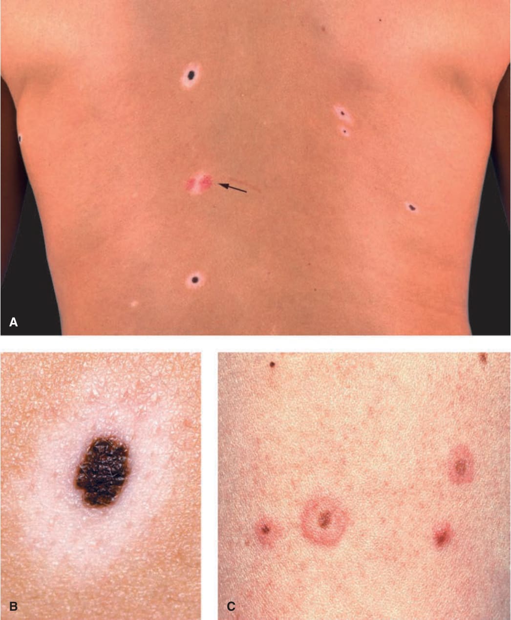
FIGURE 9-5 • (A) Halo melanocytic MN on the back of a 22-year-old female There are five halo nevi, all with a pigmented dot-like central junctional or compound MN surrounded by a hypo- or amelanotic halo. The arrow indicates one lesion where the central nevus has completely regressed; the reddish color is caused by telangiectasia. (B) Larger magnification of a halo MN. The nevus is a junctional MN (compare with Fig. 9-2) that is surrounded by a hypomelanotic almost white halo. (C) Several tan, junctional MN that are surrounded by an erythematous halo. This is the early stage of halo development but is observed only rarely. The erythematous rim will later turn white.