LEUKONYCHIA
LEUKONYCHIA
True Leukonychia. Nail matrix defect
• Total leukonychia: Usually inherited.
• Transverse leukonychia: 1- to 2-mm wide horizontal bands.
• Punctate leukonychia: Psoriasis and trauma.
• Longitudinal leukonychia: Darier disease (see Fig. 32-11).
Pseudoleukonychia. SWO (Fig. 32-20), chemical damage to nail keratin. Apparent Leukonychia. Nail bed edema, blanches with pressure to nail plate.
• Terry’s nails
• Association: Hepatic disorders.
• Findings: White discoloration obscuring lunula and extending to within 1 to 2 mm from distal edge of nail (Fig. 32-23). Involves all nails evenly.
• Half-and-Half Nail (Lindsay’s nails)
• Association: Renal disorders.
• Findings: White discoloration of proximal half of nail plate.
• Banded nails (Muehrcke lines) (see Fig. 32-33).
• Paired, narrow, and white transverse bands.
• Association: Cytotoxic chemotherapy, hypoalbuminemia.
• Findings: Bands are parallel to lunula, separated from one another, and from lunula, by strips of pink (normal) nail.
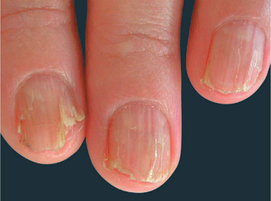
FIGURE 32-11 • Darier disease Red and white longitudinal streaks on the fingernails with V-nicking in distal portion of plate. (Reproduced with permission from Goldsmith LA, Katz SI, Gilchrest BA, et al., eds. Fitzpatrick’s Dermatology in General Medicine. 8th ed. New York, NY: McGraw-Hill; 2012.)
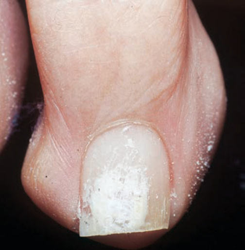
FIGURE 32-20 • Superficial white onychomycosis (SWO) The dorsal nail plate is chalky white. KOH preparation of the curetted area shows hyphae.
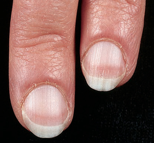
FIGURE 32-23 • Apparent leukonychia: Terry’s nails The nail plate has a white appearance, except for the distal 2 to 3 mm.
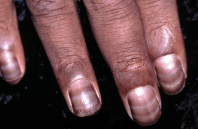
FIGURE 32-33 • Nail discoloration and transverse bands (Muehrcke lines) Period transverse bands on the fingernail in a patient with breast cancer being treated with chemotherapy (5-fluorouracil).