SEBORRHEIC DERMATITIS
SEBORRHEIC DERMATITIS ICD-10: L21.9
• A very common chronic dermatosis characterized by redness and scaling, which occurs in regions where the sebaceous glands are most active, such as the face and scalp, the presternal area, and in the body folds. Mild scalp SD causes flaking, that is, dandruff.
• Associated with a proliferation of Malassezia furfur.
• Increased incidence in Parkinson disease and in immunosuppressed patients.
Synonyms: “Cradle cap” (infants), pityriasis sicca (dandruff).
EPIDEMIOLOGY AND ETIOLOGY
AGE OF ONSET Infancy (within the first months), puberty, or most often between 20 and 50 years or older. SEX More common in males. INCIDENCE 2% to 5% of the population.
PATHOGENESIS, PREDISPOSING, AND AGGRAVATING FACTORS
The cause of seborrheic dermatitis is not fully understood, but is thought to be due to M. furfur and an abnormal immune response. There is an increase in incidence with neurologic disorders and immunosuppression.
CLINICAL MANIFESTATION
DURATION OF LESIONS Gradual onset. SEASONAL VARIATIONS Some patients are worse in winter in a dry, indoor environment. Sunlight exposure causes SD to flare in a few patients and promotes improvement of the condition in others. SKIN SYMPTOMS Pruritus is variable, often increased by perspiration. SKIN LESIONS Orange-red or gray-white skin, often with “greasy” or white dry scaling macules, papules of varying size (5 to 20 mm), or patches, rather sharply marginated (Fig. 2-23). On the scalp, there is mostly marked scaling (“dandruff”), diffuse involvement of scalp. Scattered, discrete on the face. Nummular, polycyclic, and even annular on the trunk. DISTRIBUTION AND MAJOR TYPES OF LESIONS (BASED ON LOCALIZATION AND AGE) Hairy Areas of Head Scalp, eyebrows, eyelashes, beard (follicular orifices); cradle cap: erythema and yellow-orange scales and crusts on the scalp of infants.
Face The flush (“butterfly”) areas on forehead (“corona seborrheica”), nasolabial folds, eyebrows, and glabella (Fig. 2-23). Ears: retroauricular, meatus, sticky crusts, and fissures. Trunk Simulating lesions of pityriasis rosea or pityriasis versicolor; yellowish-brown patches over the sternum common. Body Folds Axillae, groins, anogenital area, submammary areas, umbilicus, and diaper area in infants (Fig. 2-24)—presents as a diffuse, exudative, sharply marginated, brightly erythematous eruption; erosions and fissures common. Genitalia Often with yellow crusts and psoriasiform lesions.
DIAGNOSIS/DIFFERENTIAL DIAGNOSIS
Made on clinical criteria. RED SCALY PLAQUES Common Mild psoriasis vulgaris (sometimes may be indistinguishable), impetigo (rule out by smears for bacteria), dermatophytosis, pityriasis versicolor, intertriginous candidiasis (rule out dermatophytes and yeasts by KOH), subacute lupus erythematosus (rule out by biopsy), “seborrheic” papules in secondary syphilis (rule out Treponema pallidum by dark field); syphilis serology. Rare Langerhans cell histiocytosis (occurs in infants, often associated with purpura), acrodermatitis enteropathica, zinc deficiency, pemphigus foliaceus, glucagonoma syndrome.
LABORATORY STUDIES
DERMATOPATHOLOGY Focal parakeratosis, with few neutrophils, moderate acanthosis, spongiosis (intercellular edema), and nonspecific inflammation of the dermis. Neutrophils at the tips of the dilated follicular openings, which appear as crusts/scales.
COURSE AND PROGNOSIS
The condition improves in the summer and flares in the fall. Recurrences and remissions, especially on the scalp, may be associated with alopecia in severe cases. Infantile and adolescent SD disappears with age. Seborrheic erythroderma may occur. Seborrheic erythroderma with diarrhea and failure to thrive in infants (Leiner disease) is associated with a variety of immunodeficiency disorders including defective yeast opsonization, C3 deficiency, severe combined immunodeficiency,
hypogammaglobulinemia, and hyperimmunoglobulinemia.
MANAGEMENT
Requires initial therapy followed by chronic maintenance therapy.
Initial Topical Therapy
SCALP Adults Shampoos containing selenium sulfide, zinc pyrithione, and/or tar. 2% ketoconazole shampoo is very effective; lather can be used on face and chest during shower. Low-potency glucocorticoid solution, lotion,
or gels following a medicated shampoo (ketoconazole or tar) for more severe cases. Topical calcineurin inhibitors can also be beneficial. Infants For cradle cap, removal of crusts with warm olive oil compresses, followed by baby shampoo, 2% ketoconazole shampoo, and application of 1% to 2.5% hydrocortisone cream, 2% ketoconazole cream, and topical calcineurin inhibitors. FACE AND TRUNK Ketoconazole shampoo, 2%. Glucocorticoid cream and lotions. 2% ketoconazole cream, topical calcineurin inhibitors. EYELIDS Gentle removal of the crusts in the morning with a cotton ball dipped in diluted baby shampoo.
INTERTRIGINOUS AREAS Ketoconazole, 2%. Low potency topical glucocorticoid, topical calcineurin inhibitors.
Systemic Therapy In severe cases, oral fluconazole can be considered.
Maintenance Therapy Ketoconazole 2% shampoo, tar shampoos, and ketoconazole cream are effective. 1% to 2.5% hydrocortisone cream daily will work, but patients should be monitored for signs of atrophy; 1% pimecrolimus cream and 0.03%/0.1% tacrolimus ointments are also safe and effective.
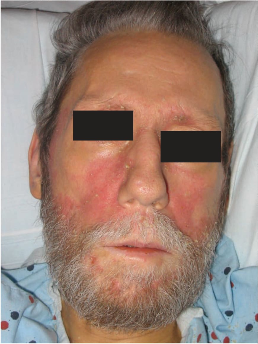
FIGURE 2-23 • Seborrheic dermatitis of face: adult type Erythematous patches with “greasy” scale on the eyebrows, nasolabial folds, and beard area.
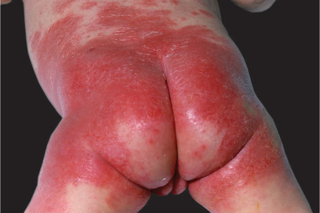
FIGURE 2-24 • Seborrheic dermatitis: infantile type Erythema scales and crusting in the diaper region of an infant. This is difficult to distinguish in the diaper region from psoriasis and Candida has to be ruled out by KOH.