HAILEY–HAILEY DISEASE (FAMILIAL BENIGN PEMPHIGUS)
HAILEY–HAILEY DISEASE (FAMILIAL BENIGN PEMPHIGUS) ICD-10: Q82.8
• Hailey–Hailey disease, or familial benign pemphigus, is a rare genodermatosis with dominant inheritance that is classically described as a blistering disorder but actually presents as an erythematous, erosive, oozing condition with cracks and fissures localized to the nape of the neck, axillae (Fig. 5-6).
• Inframammary regions, inguinal folds, and scrotum are major sites of involvement.
• Individual lesions consist of microscopically small flaccid vesicles on an erythematous background that soon turn into eroded plaques with the described, highly characteristic, fissured appearance (Fig. 5-6). Crusting, scaling, and hypertrophic vegetative lesions occur.
• The underlying pathologic process is acantholysis whereby the fragility of the epidermis results from a defect in the adhesion complex between desmosomal proteins and tonofilaments.
• The genetic abnormality lies in ATP2CI, which encodes an ATP-powered calcium pump.
• Onset is usually between the third and fourth decades.
• Crusting, scaling, and hypertrophic vegetative growths may occur.
• Histology explains the clinical appearance as epidermal cells lose their coherence with acantholysis throughout the epithelium, giving the appearance of a dilapidated brick wall.
• Colonization of the lesions, particularly by Staphylococcus aureus and candida, is a trigger for further acantholysis and maintenance of the pathologic process.
• Treatment rests on antimicrobial therapy, administered both topically and systemically; systemically, the tetracyclines seem to work better than most. Topical glucocorticoids depress the anti-inflammatory response and accelerate healing. In severe cases, dermabrasion or carbon dioxide laser vaporization leads to healing with scars, which are resistant to recurrences. Systemic retinoids and low-dose naltrexone may also be considered. The condition becomes less troublesome with age.
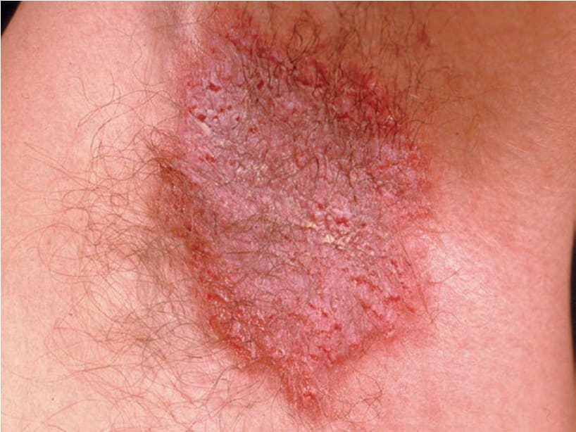
FIGURE 5-6 • Hailey–Hailey disease This 46-year-old male had oozing lesions on both armpits, occasionally in the groins and nape of the neck for several years, which became worse during summer months. Father and sister have similar lesions. Lesions wax and wane, are painful, and show typical cracks and fissures within a partially erosive erythematous plaque.