BLUE NEVUS
BLUE NEVUS ICD-10: D22. L42
• A blue nevus is an acquired, firm, dark-blue to gray-to-black, sharply defined papule or nodule representing a localized proliferation of melanin-producing dermal melanocytes.
• Three types: common blue nevus, cellular blue nevus, combined melanocytic nevus/ blue nevus.
• Blue nevi and combined melanocytic/blue nevi are benign. Cellular blue nevi are larger with a very rare tendency to become malignant.
• Ectopic accumulation of melanin-producing melanocytes; derived from melanoblasts arrested during migration from neural crest.
• Papules, nodules, blue-gray, blue-black, <10 mm in diameter (Figs. 9-6 and 9-7A). Cellular blue nevi larger (>1 cm) and irregular (Fig. 9-7B).
• Differential diagnosis: Dermatofibroma, glomus tumor, nodular or metastatic melanoma, traumatic tattoo, pigmented BCC.
• If in doubt of malignancy, excision. Cellular blue nevi should also be excised.
• Synonyms: Blue neuronevus, dermal melanocytoma.
B A
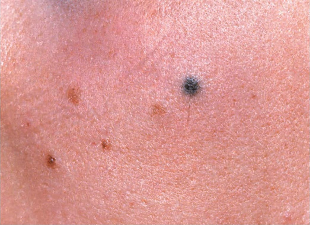
FIGURE 9-6 • Blue nevus There are four tan junctional MN and one bluish-black round lesion on the cheek of a 17-year-old girl. In contrast to the junctional MN, the blue nevus is palpable with a relatively high consistency, and upon dermoscopy will appear as an ill-defined uniformly bluish lesion deep in the dermis.
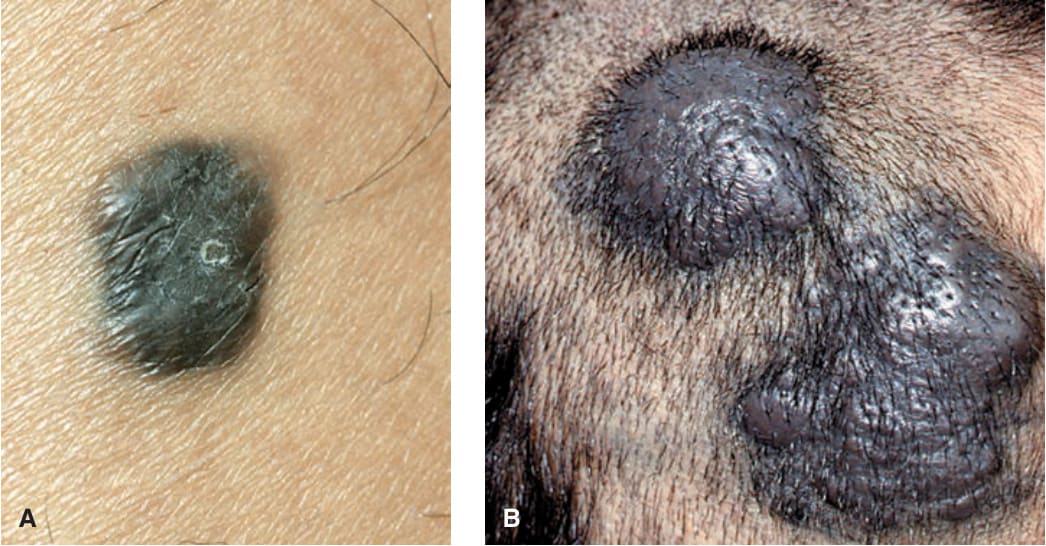
FIGURE 9-7 • Blue nevus and cellular blue nevus (A) This blue nevus has regular borders but is not circular and is solidly blue–black in color. The epidermis is smooth, indicating that the lesion is in the dermis. The consistency is increased and the margins are well defined. Differential diagnosis must include nodular melanoma. (B) This cellular blue nevus appeared as two large, bluish-black nodules on the scalp. After excision, histology showed that they were contiguous and thus represented one single lesion. Cellular blue nevi are much larger and should always be excised to rule out melanoma, which, albeit rarely, can develop in these lesions.