SPITZ NEVUS
SPITZ NEVUS ICD-10: D22.L30
• Spitz nevus is a benign, dome-shaped, hairless, small (<1 cm in diameter) nodule, most often pink, red, or tan (Fig. 9-9A). There is often a history of recent rapid growth.
• Incidence is 1.4:100,000 (Australia). Most commonly arises in first two decades. They are dome-shaped, firm, and smooth papules/nodules. They can be pink-red (Fig. 9-9A), tan, brown, dark brown, or even black (Fig. 9-9B); usually distributed on the face and legs.
• Differential diagnosis includes all pink, tan, or darkly pigmented papules: pyogenic granuloma, hemangioma, molluscum contagiosum, juvenile xanthogranuloma, mastocytoma, dermatofibroma, melanocytic nevi, and nodular melanoma.
• Dermatopathology : Hyperplasia of the epidermis and melanocytes and dilation of capillaries. Admixed large epithelioid cells, large spindle cells with abundant cytoplasm, and occasional mitotic figures.
• Histologic examination must be done to confirm the clinical diagnosis. Excision in its entirety is important because the condition recurs in 10% to 15% of all cases in lesions that have not been excised completely. Spitz nevi are benign, but there can be a histologic similarity to melanoma and the histopathologic diagnosis requires the help of an experienced dermatopathologist.
• Spitz nevi do not usually involute, though some may persist.
• Synonyms: Pigmented and epithelioid spindle-cell nevus. Decades ago, these were called “juvenile melanoma.”
A B
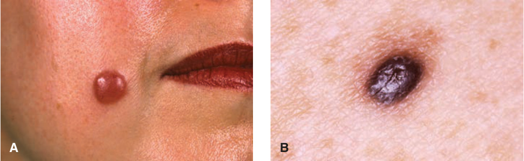
FIGURE 9-9 • Spitz nevus (A) Pink dome-shaped nodule on the cheek of a 25-year-old woman, developing abruptly within the previous 12 months; the lesion can be mistaken for a hemangioma. (B) Pigmented Spitz nevus (also called Reed nevus). A black papule surrounded by a tan macular region developed within a few months on the back of a young female; as such a lesion cannot be distinguished from a nodular melanoma, the lesion was excised and the diagnosis confirmed histologically.