CONGENITAL DERMAL MELANOCYTOSIS
CONGENITAL DERMAL MELANOCYTOSIS ICD-10: D22.505
• These congenital gray–blue macular lesions are characteristically located on the lumbosacral area (Fig. 9-10) but can also occur on the back, scalp, or anywhere on the skin. There is usually a single lesion, but rarely, several truncal lesions can be present at birth (Fig. 9-11).
• The underlying pathology is dispersed spindle-shaped melanocytes within the dermis. It is believed that these ectopic melanocytes represent pigment cells that have been interrupted in their migration from the neural crest to the epidermis.
• Congenital dermal melanocytosis may disappear in early childhood, in contrast to nevus of Ota (see Fig. 9-12).
• These lesions are found in most Asian and Brown newborns. It presents in less than 10% of white infants.
• No melanomas have been reported to occur in these lesions.

FIGURE 9-10 • Congenital dermal melanocytosis A large gray–blue macular lesion involving the entire lumbosacral and gluteal area and the left thigh in a baby from Sri Lanka. Although these lesions are common in Asians, the parents of this baby were alarmed because the lesion was so large.
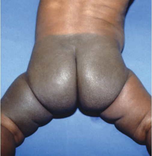
FIGURE 9-11 • Congenital dermal melanocytosis Multiple, ill-defined, bluish lesions are scattered on the back of this Japanese child. They were present at birth. Most of these lesions disappeared later in childhood.
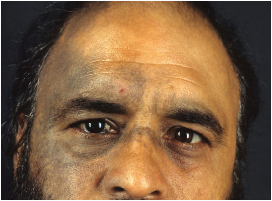
FIGURE 9-12 • Nevus of Ota There is an ill-defined, mottled, dusky, gray to bluish hyperpigmentation in the regions supplied by the first and second branches of the right trigeminal nerve. The lesion was unilateral and there was also hyperpigmentation of the sclera and eyelids.