PORT-WINE STAIN (PWS)
PORT-WINE STAIN (PWS) ICD-10: Q82.510
• A PWS is an irregularly shaped, red or violaceous, macular capillary malformation that is present at birth and never disappears spontaneously.
• It is common (0.3% of newborns); the malformation is usually confined to the skin.
• May be associated with vascular malformations in the eye and leptomeninges (Sturge–Weber syndrome [SWS]).
• Synonym: Nevus flammeus.
SKIN LESIONS Macular (Fig. 9-19) with varying hues of pink to purple. Large lesions follow a dermatomal distribution, usually unilateral (85%), although not always. Most commonly in the face, in the distribution of the trigeminal nerve (Fig. 9-19), and usually the superior and middle branches; mucosal involvement of conjunctiva and mouth may occur. May also involve other sites. With increasing age of the patient, papules or rubbery nodules (Fig. 9-20) cause significant disfigurement.
Clinical Variant Nevus simplex (“stork bite,” “salmon patch,” “angels kiss”) occurs in ∼50% infants. Lesions occur on the eyelid, glabella, and neck. Most resolve in 1 to 2 years, except for those on the neck, which persist.
HISTOPATHOLOGY
Reveals ectasia of capillaries and no proliferation of endothelial cells. GLUT-1 immunoreactivity is negative.
COURSE AND PROGNOSIS
Port-wine stain (PWS) does not regress spontaneously. The area of involvement tends to increase in proportion to the size of the child. In adulthood, PWS usually become raised
with papular and nodular areas that can cause significant cosmetic disfigurement (Fig. 9-20).
MANAGEMENT
Treatment with pulsed dye lasers is highly effective.
SYNDROMIC CAPILLARY MALFORMATIONS
Sturge–Weber syndrome (SWS) is the association of PWS in the trigeminal distribution with vascular malformations in the eye and leptomeninges, and superficial calcifications of the brain. May be associated with contralateral hemiparesis, muscular hemiatrophy, epilepsy, mental retardation, and glaucoma and ocular palsy. Characteristic calcifications of vascular malformations or localized linear calcification along cerebral convolutions at x-ray. CT scan should be done. It should, however, be noted that PWS with trigeminal distribution is common and does not necessarily indicate the presence of SWS. Klippel–Trénaunay– Weber syndrome may have an associated PWS overlying the deeper vascular malformation of soft tissue and bone. PWS on the midline back may be associated with an underlying AVM of the spinal cord.
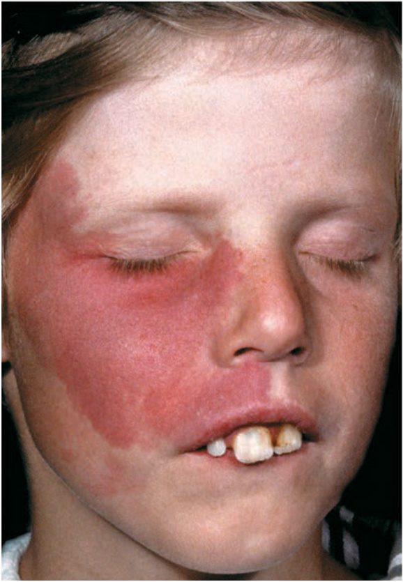
FIGURE 9-19 • Port-wine stain Sharply marginated, port-wine red macule occurring in a distribution of the second branch of the trigeminal nerve in a child.
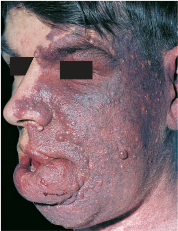
FIGURE 9-20 • Port-wine stain With increasing age, the color deepens and papular and nodular vascular lesions develop within the previously macular lesion, causing progressively increasing disfigurement.