ANGIOKERATOMA
ANGIOKERATOMA ICD-10: code according to type (see text)
• The term angio (“blood vessel”) keratoma would imply a vascular tumor with keratotic elements. But, capillaries and postcapillary venules are packed into the papillary body just beneath and bulging into the epidermis, leading to hyperkeratosis. This and the fact that the lumina are usually at least partially thrombosed impart a firm consistency to the lesions.
• Angiokeratomas are dark violaceous to black, often keratotic papules or small plaques that are hard upon palpation and cannot be compressed by diascopy (Fig. 9-24).
• Angiokeratoma can appear as a solitary lesion (solitary angiokeratoma). The most important differential diagnosis is a small nodular or superficial spreading melanoma (Fig. 9-24).
• The most common is angiokeratoma of Fordyce (ICD10:D29.420); this disease involves the scrotum and vulva; the lesions are multiple papules (≤4 mm) that are dark red to black in color and present in quite large numbers (Fig. 9-25).
• Angiokeratoma of Mibelli (ICD-10:D23.L74) comprises pink to dark red and even black papules that occur on the elbows, knees, and dorsa of the hands. This autosomal-dominant disease is rare and occurs in young females.
• Angiokeratoma corporis diffusum (Fabry disease) (ICD10:E75.250), an X-linked recessive disease, is an inborn error of metabolism in which there is a deficiency of α-galactosidase A leading to an accumulation of neutral glycosphingolipid ceramide trihexoside in endothelial cells, fibrocytes, and pericytes in the dermis, heart, kidneys, and autonomic nervous system. Lesions are numerous, dark red, punctate, and tiny (<1 mm) (Fig. 9-26), located on the lower half of the body: lower abdomen, genitalia, and buttocks, although lesions may also occur on the lips. The homozygous males also have symptoms related to involvement of other organ systems: acroparesthesias, excruciating pain, transient ischemic attacks, and myocardial infarction. Heterozygous females may have corneal opacities. Fabry disease is very rare but serious.
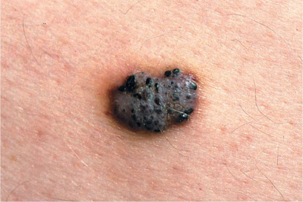
FIGURE 9-24 • Angiokeratoma: solitary This black, firm lesion with a pebbled surface immediately sparks the suspicion of superficial spreading melanoma. It is noncompressible, but dermoscopy reveals the typical lacunae of thrombosed vascular spaces. Nonetheless, such lesions should be excised.
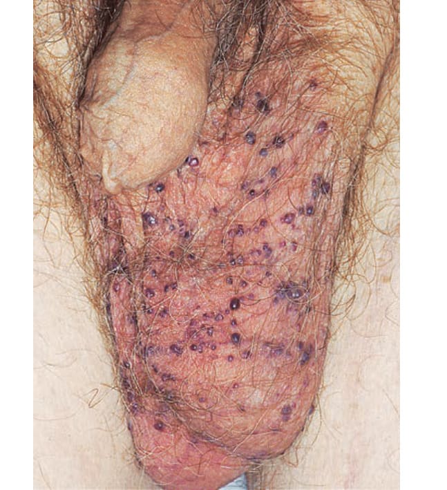
FIGURE 9-25 • Angiokeratoma of Fordyce Reddish, violaceous, and black papules on the scrotum. They blanch upon diascopy and this verifies the diagnosis. Note: Thrombosed angiokeratomas do not blanch.
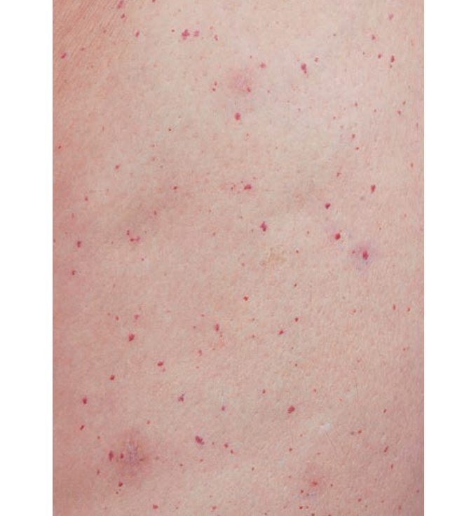
FIGURE 9-26 • Angiokeratoma corporis diffusum (Fabry disease) Numerous red, punctate lesions on the lower flank.