DERMATOHELIOSIS (“PHOTOAGING”)
DERMATOHELIOSIS (“PHOTOAGING”) ICD-10: L57.91
• Skin changes secondary to continuous long-term ultraviolet radiation exposure.
• It occurs in persons with SPT I to III and in persons with SPT IV who have had heavy cumulative exposure to sunlight, such as lifeguards and outdoor workers, over a lifetime. Most often in persons >40 years.
• Severity depends on the duration and intensity of sun exposure and skin phototype.
• Skin lesions: A combination of atrophy (of epidermis), hypertrophy (of papillary dermis caused by elastosis), telangiectases, spotty depigmentation and hyperpigmentation, and spotty hyperkeratosis in light-exposed areas. Skin appears wrinkled, leathery, and “prematurely aged” (Fig. 10-20). Both fine, cigarette paper-like, and deep furrow-like wrinkling; skin is waxy, papular with a yellowish hue, and both glistening and rough (Fig. 10-21). Telangiectasia and bruising (senile purpura) caused by fragility of small vessels. Macular hyperpigmentations: Solar lentigines (see the following discussion); macular hypopigmentations: guttate hypomelanosis, <3 mm in diameter, on the extremities. Comedones, particularly periorbital (termed Favre– Racouchot disease) (Fig. 10-22), particularly in cigarette smokers. Individuals invariably have actinic keratoses.
• Distribution: Exposed areas, particularly face, periorbital and perioral areas, and scalp (bald males). Nuchal area: Cutis rhomboidalis (“red neck”) with rhomboidal furrows; lower arms, dorsa of the hands.
• Current management is to prevent skin cancers and the development of dermatoheliosis with the use of protective sunblocks, a change of behavior in the sun, and the use of topical chemotherapy (tretinoin) that reverses some of the skin changes.
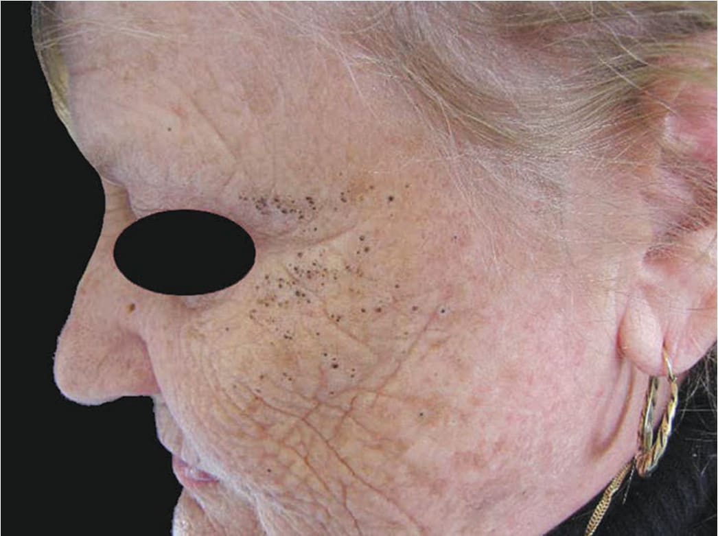
FIGURE 10-20 • Dermatoheliosis Severe deep wrinkling. The skin appears waxy, papular with a yellowish hue (solar elastosis). This 68-year-old female mountain farmer lived at an altitude of 1500 m and had been working outdoors all her life. In addition, she was a heavy smoker and now has multiple black comedones in the zygomatic regions (Favre–Racouchot disease). (Used with permission from Dr. Gudrun Ratzinger.)
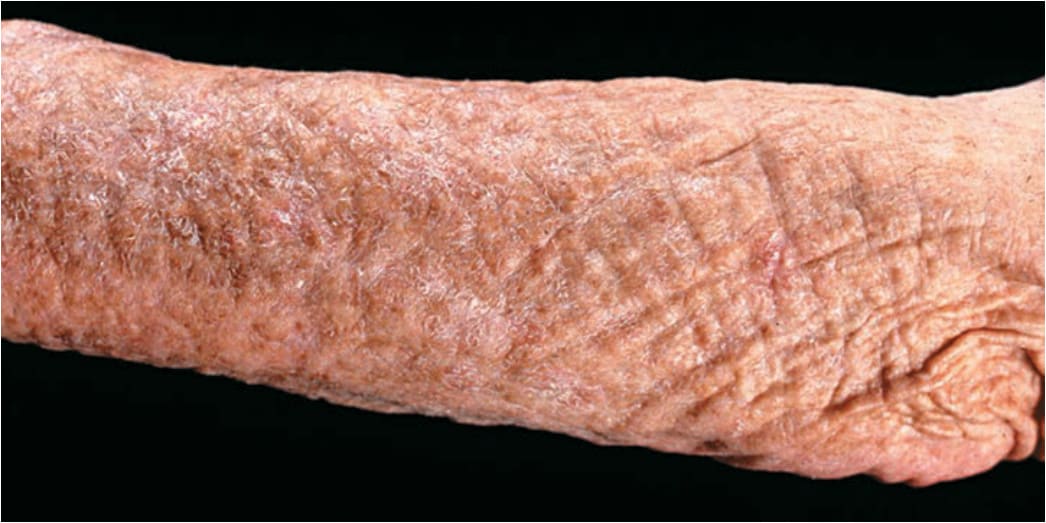
FIGURE 10-21 • Severe dermatoheliosis on the forearm of a 70-year-old female farmhand The skin is waxy, deeply wrinkled, and dry. Multiple solar keratoses have been removed from this arm by cryotherapy.
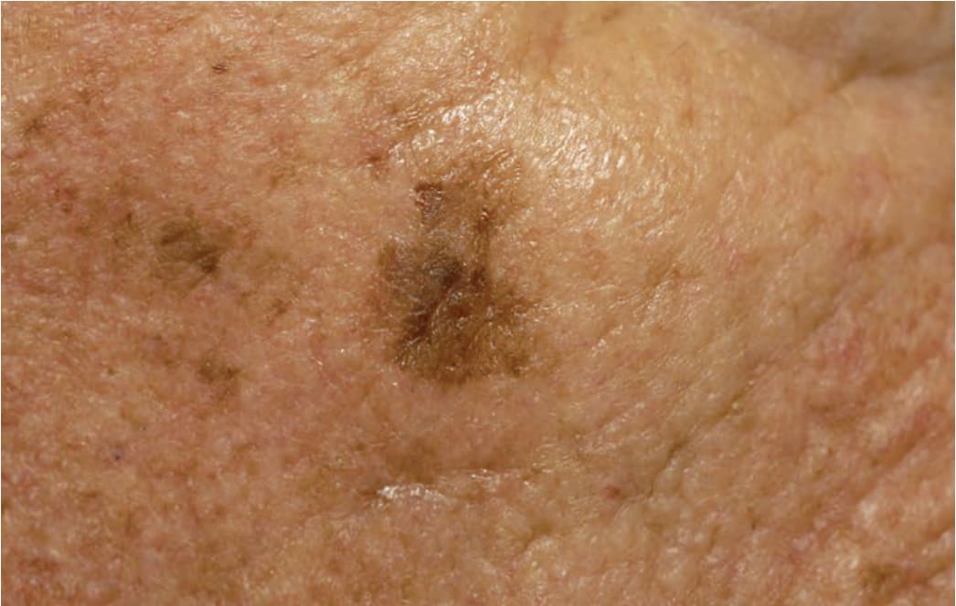
FIGURE 10-22 • Dermatoheliosis: solar lentigines Multiple, very small to large (2 cm), variegated, tan-to-darkbrown macules on the cheek. Solar lentigines are not the same as ephelides (freckles). They do not fade in the winter as freckles do. In contrast to the sharply marginated solar lentigines caused by an acute sunburn that have roughly the same size shown in Figure 10-24, the solar lentigines shown here are of different sizes and partially ill defined and confluent, which is characteristic of chronic cumulative solar damage. Note waxy thickening of skin and creases of dermatoheliosis.