SOLAR LENTIGO
SOLAR LENTIGO ICD-10: L81.416
• Solar lentigo is a circumscribed 1- to 3-cm brown macule resulting from a localized proliferation of melanocytes resulting from acute or chronic exposure to sunlight.
• Onset usually >40 years.
• Multiple lesions usually arise in sun-exposed sites. Most common in patients with SPT I and II.
• Macules, 1 to 3 cm, and as large as 5 cm. Light yellow, light brown, or dark brown; variegated mix of brown (Fig. 10-22). Round, oval, with slightly irregular border, and ill defined. Scattered, discrete lesions, stellate, sharply defined, and roughly the same size after acute sunburn (Fig. 10-23) or overdosage of PUVA.
• Distribution. Exclusively exposed areas: forehead, cheeks, nose, dorsa of the hands and forearms, upper back, chest, and shins.
• Differential diagnosis: “Flat,” acquired, brown lesions on the exposed skin of the face, which may on cursory examination appear to be similar and have distinctive features: solar lentigo, freckles, seborrheic keratosis, spreading pigmented actinic keratosis (SPAK), and lentigo maligna (LM).
• Cryosurgery or laser surgery is effective.
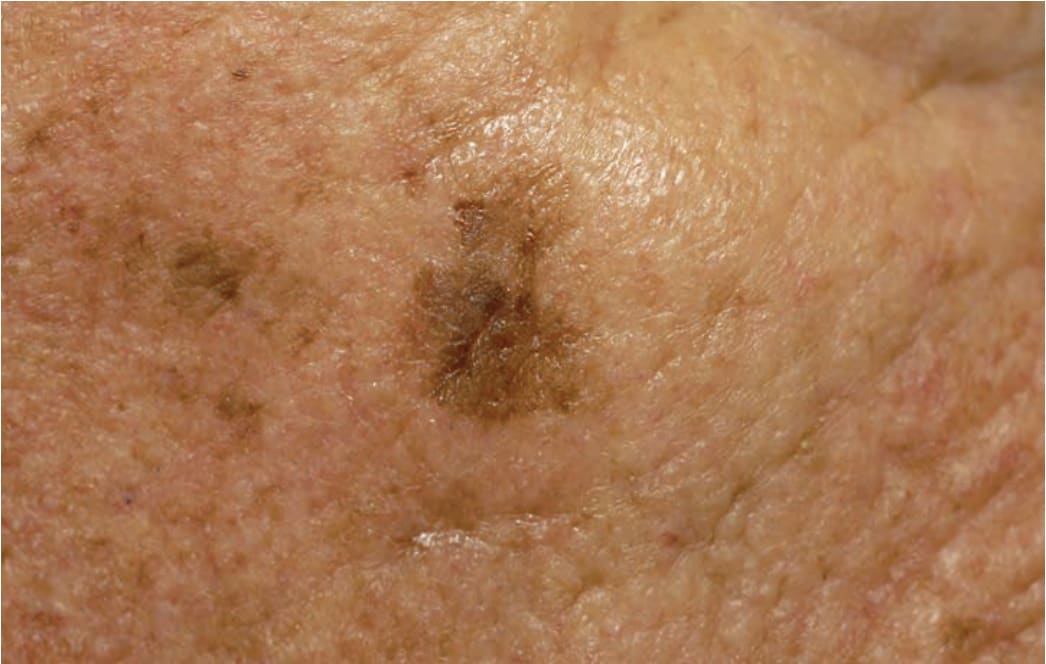
FIGURE 10-22 • Dermatoheliosis: solar lentigines Multiple, very small to large (2 cm), variegated, tan-to-darkbrown macules on the cheek. Solar lentigines are not the same as ephelides (freckles). They do not fade in the winter as freckles do. In contrast to the sharply marginated solar lentigines caused by an acute sunburn that have roughly the same size shown in Figure 10-24, the solar lentigines shown here are of different sizes and partially ill defined and confluent, which is characteristic of chronic cumulative solar damage. Note waxy thickening of skin and creases of dermatoheliosis.
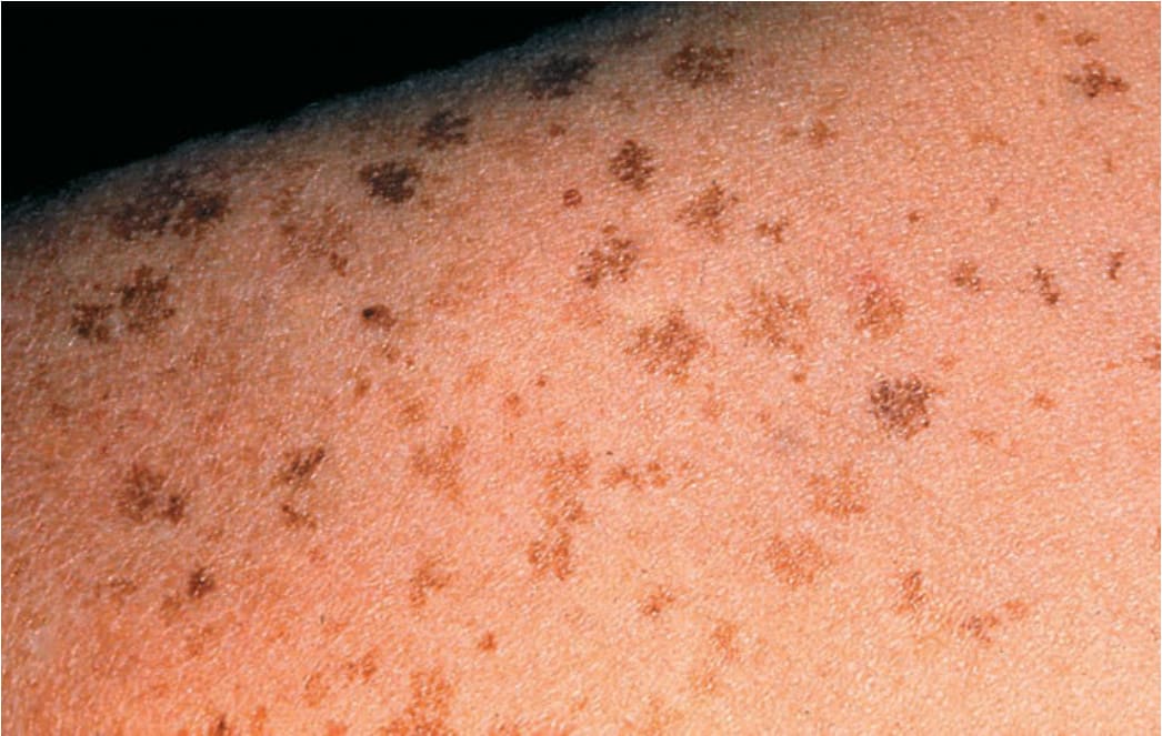
FIGURE 10-23 • Dermatoheliosis: solar lentigines Multiple stellate brown macules on the shoulder occurred after a sunburn. They are all of about the same size and sharply marginated, which is characteristic of sunburn-induced solar lentigines.