RADIATION DERMATITIS
RADIATION DERMATITIS ICD-10: L58
• Radiation dermatitis is defined as skin changes resulting from exposure to ionizing radiation.
• Reversible effects are pain, erythema, epilation, suppression of sebaceous glands, and pigmentation (lasting for weeks to months to years).
• Irreversible effects are atrophy, sclerosis, telangiectasias, ulceration, and radiation-induced cancers.
Type of Exposure Result of therapy (for cancer, formerly also used for acne and psoriasis, and fungal infections of the scalp in children), accidental, or occupational (e.g., formerly, in dentists). The radiation causing radiodermatitis includes superficial and deep x-ray radiation, electron beam therapy, and grenz-ray therapy. It is a prevailing myth that grenz rays are “soft” and not carcinogenic; squamous cell carcinoma (SCC) can appear from >5,000 cGy of grenz rays.
Types of Reactions
ACUTE Temporary erythema that lasts 3 days and then persistent erythema, which reaches a peak in 2 weeks and is painful; pigmentation appears around day 20; a late erythema can also occur beginning on day 35 to 40, and usually lasts 2 to 3 weeks. Massive reactions lead to blistering, erosions (Fig. 10-25), and ulceration, also painful; may occur as recall phenomenon. Permanent scarring may result.
CHRONIC After fractional but relatively intensive therapy with total doses of 3000 to 6000 rad, there develops an epidermolytic reaction in 3 weeks. This is repaired in 3 to 6 weeks, but scars and hypopigmentation develop; there is loss of all skin appendages and atrophy of the epidermis and dermis. During the next 2 to 5 years, the atrophy increases (Fig. 10-26); there is hyper- and hypopigmentation (poikiloderma), telangiectasia (Figs. 10-26, 10-27 and 10-28). Necrosis and painful ulceration (Fig. 10-29) are rare but occur in accidental exposure or error in dose. Necrosis is leathery, yellow, and adherent and surrounding skin are extremely painful (Fig. 10-29). Ulcerations have a very poor tendency to heal and usually
require surgical intervention. Lastly, there may be radiation keratoses and squamous cell carcinoma (Fig. 10-30). NAILS Longitudinal striations (Fig. 10-30B) show thickening, dystrophy.
COURSE, PROGNOSIS, AND MANAGEMENT
Chronic radiation dermatitis is permanent, progressive, and irreversible. SCC may develop in 4 to 40 years (Fig. 10-30), with a median of 7 to 12 years. Tumors metastasize in about 25%; despite extensive surgery (excision, grafts, etc.), the prognosis is poor, and recurrences are common. Angiosarcoma can also occur.
A B
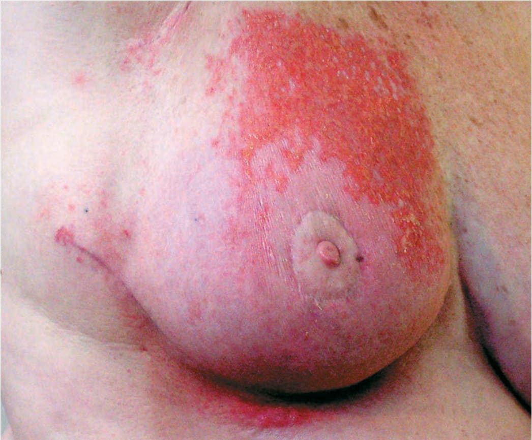
FIGURE 10-25 • Radiation dermatitis: acute, recall phenomenon This patient had breast cancer. She had a lumpectomy, methotrexate, and x-ray therapy and developed painful erythema and erosions at the irradiated site.
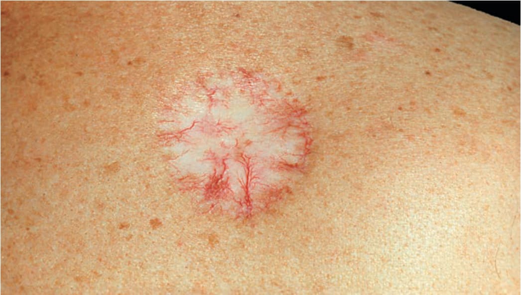
FIGURE 10-26 • Radiation dermatitis: chronic There is sclerosis combined with atrophy and telangiectasia. This is the result of the irradiation of an infantile hemangioma in infancy.
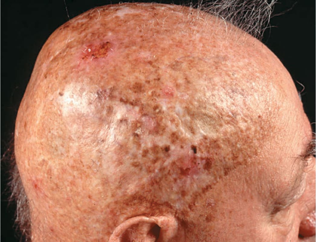
FIGURE 10-27 • Radiation dermatitis: chronic There is poikiloderma (brown: hyperpigmentation; white: hypopigmentation; red: telangiectasia) combined with atrophy and sclerosis. Hairs are absent. These massive skin changes are the result of overdosed irradiation that the patient received as a child for fungal infection of the scalp. He is a candidate for SCC in the future.
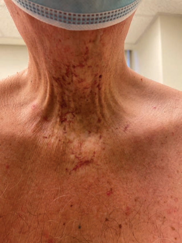
FIGURE 10-28 • Radiation dermatitis: chronic Note the dilated telangiectasias and focal sclerosis in a patient previously radiated for laryngeal carcinoma.
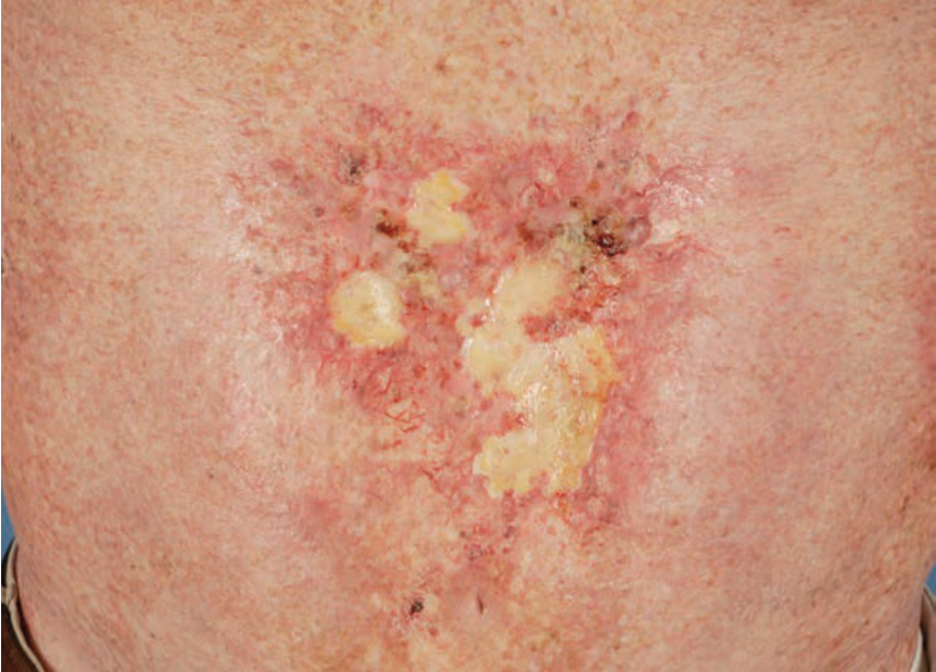
FIGURE 10-29 • Radiation dermatitis: chronic An area of severe poikiloderma with telangiectasias and irregular areas of necrosis that is leathery, yellowish-white, and tightly adherent. The lesion is extremely painful. Occurred after repeated electron beam radiations for mycosis fungoides.
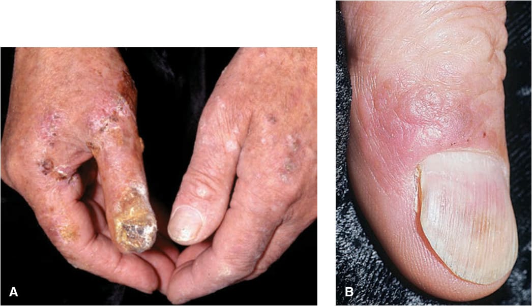
FIGURE 10-30 • Radiation-induced squamous cell carcinoma (A) These are the hands of an elderly radiologist who decades ago had disregarded precautionary measures and hardly wore gloves doing fluoroscopic work. There are multiple x-ray keratoses; the hyperkeratotic lesion on the right thumb has destroyed the nail and represents x-ray– induced SCC. (B) Nail changes in the site of radiation exposure. Note the linear striations resulting from damage to the nail matrix. At the nailfold and extending proximally on the thumb, there is an irregular erythematous plaque that represents mostly SCC in situ but also focally invasive SCC.