OCULOCUTANEOUS ALBINISM
OCULOCUTANEOUS ALBINISM ICD-10: E70.390
• Classification, see Table 13-1.
• Prevalence estimated 1:20,000 OCA1 and OCA2 account for 40% to 50%.
• Mutations in the tyrosinase gene are responsible for deficient tyrosinase activity in melanocytes (Table 13-1).
• Present at birth.
• Skin varied depending on type. “Snow white,” creamy white (Fig. 13-7; Table 13-1), light tan.
• Hair: White (tyrosinase negative; Fig. 13-7A); yellow, cream, light brown (tyrosinase positive); red, platinum (Table 13-1).
• Eyes: Nystagmus, reduction of visual activity, iris translucency (Fig. 13-7B), decreased retinal pigment, foveal hyperplasia, and strabismus.
• Dermatopathology: Melanocytes are present but tyrosinase reduced depending on type.
• Molecular testing. Available to classify specific gene alterations.
• Significance: Reduction of visual activity; development of dermatoheliosis, and skin cancer without sun protection. Especially important for albinos living in Africa (Fig. 13-8).
• Management: No treatment available. Albinos should be under care of an ophthalmologist (vision problems) and a dermatologist (sun protection and detection of skin cancer).
• National volunteer group of albinos (in the United States: NOAH—National Organization for Albinism and Hypomelanosis ([Noah of the Old Testament was alleged to be an Albino]).
Type Subtypes Gene Locus Includes Clinical Findings
OCA1 OCA1A OCA1B TYR TYR Tyrosinase-negative OCA Minimal pigment OCA Platinum OCA
White hair and skin, eyes (pink at birth → blue) White to near-normal skin and hair pigmentation Yellow (pheomelanin) hair, light red or brown hair Temperature-sensitive OCA Autosomal-recessive OCA (some) May have near-normal pigment but not in axilla
Yellow OCA
OCA2 P Tyrosinase-positive OCA Brown OCA Yellow hair, skin “creamy” white (Africa) Light brown/tan skin (Africa)
OCA3 TYRP1 Autosomal-recessive OCA (some) Rufous OCA Red and red-brown skin and brown eyes (Africa) Similar to OCA2 phenotype
OCA4 MATP
HPS HPS Hermansky–Pudlak syndrome Types 1–8 Skin/hair as in OCA1A or OCA1B or OCA2, bleeding diathesis (Puerto Rico)
CHS LYST Chédiak–Higashi syndrome Silver hair/hypopigmentation/serious medical problems
OA1 OA1 X-linked OA Normal pigmentation of skin and hair
LYST, lysosome trafficking; MATP, membrane-associated transporter protein; OA, ocular albinism; OCA, oculocutaneous albinism; P, pink protein; TYR, tyrosinase; TYRP1, tyrosinase-related protein 1. Source: Modified with permission from Bahadoran P, et al. Albinism. In: Freedberg IM, Eisen AZ, Wolff K, et al., eds. Fitzpatrick’s Dermatology in General Medicine. 6th ed. New York, NY: McGraw-Hill; 2003.
A B
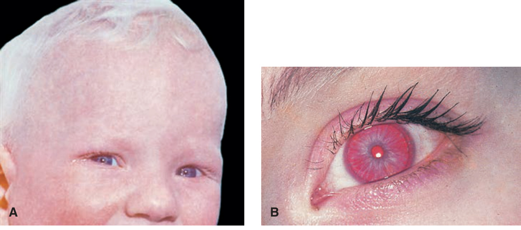
FIGURE 13-7 • (A) Oculocutaneous albinism White skin, white eyelashes, eyebrows, and scalp hair. The irises appear translucent. Heme pigment gives the face a pinkish hue. There is squinting resulting from photophobia and nystagmus. (B) Iris translucency is a sine qua non in all types of oculocutaneous albinism, even in those patients in whom the iris is brown. The iris is rarely pink except in infants, and the diagnosis of albinism depends on the detection of iris translucency. This is best done in a dark room with a pointed flashlight placed on the sclera.
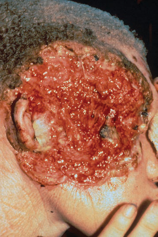
FIGURE 13-8 • Squamous cell carcinoma in an Albino from Tanzania This 32-year-old African was completely white and thus unprotected from solar exposure. The carcinoma started at the age of 28 and has destroyed most of the right face including the eye. There were smaller tumors on the left side of the face and the hands and lower arms. The patient succumbed to metastatic carcinoma.
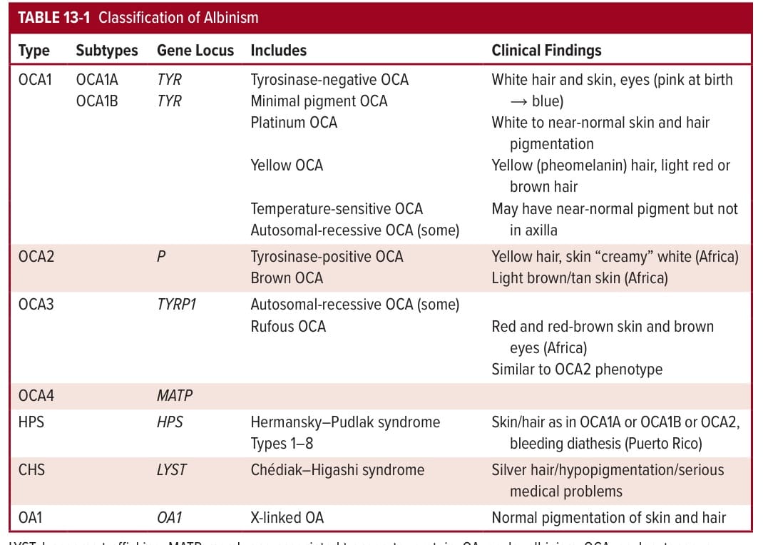
TABLE 13-1 Classification of Albinism