SARCOIDOSIS
SARCOIDOSIS ICD-10: D86
• A systemic granulomatous disease of unknown cause.
• Primarily affecting the lungs (bilateral lymphadenopathy and pulmonary infiltration).
• Skin: Papules, translucent yellow-red with apple jelly appearance on diascopy; nodules and bluish-red plaques.
• Often localizes in scars.
• Histologically, noncaseating, and “naked” granulomas.
• Erythema nodosum is the most common nonspecific lesion in the skin in early sarcoidosis; it suggests a good prognosis.
EPIDEMIOLOGY
AGE OF ONSET Under 40 years (range 12 to 70 years). SEX Equal incidence in males and females. RACE The disease occurs worldwide, frequently in Scandinavia. All races. In the United States and South Africa, much more frequent in dark skinned ethnicities. OTHER FACTORS Etiology unknown. The disease can occur in families.
CLINICAL MANIFESTATION
Onset of lesions: Days (presenting as acute erythema nodosum) or months (presenting as asymptomatic sarcoidal papules or plaques on the skin or pulmonary infiltrate discovered on routine chest radiography). Constitutional symptoms such as fever, fatigue, weight loss, and arrhythmia.
SKIN LESIONS Earliest lesions are skin-colored or brownish papules, occurring periorificially on the face (Fig. 14-66). Brownish or purple infiltrated plaques that may be annular, polycyclic, serpiginous, and occur mainly on the extremities, buttocks, and trunk. Central clearing with slight atrophy may occur. Occasionally, nodules, firm, purple or brown, may arise on the face, trunk, or extremities, particularly the hands. Lupus pernio: Diffuse, violaceous, soft doughy infiltrations on the nose, cheeks (Fig. 14-67), or earlobes. Swelling of individual digits because of osteitis cystica (Fig. 14-68). Sarcoidosis tends to infiltrate old scars, which then exhibit translucent purple-red or yellowish papules or nodules (Fig. 14-69A). Note: On blanching with glass slide, all cutaneous lesions of sarcoidosis reveal “apple jelly” semitranslucent
yellowish brown color (Fig. 14-69B). On the scalp, sarcoidosis may cause scarring alopecia (see Section 31). SYSTEMS REVIEW Enlarged parotids, pulmonary infiltrates, cardiac dyspnea, neuropathy, uveitis, kidney stones. Löfgren syndrome: erythema nodosum, fever, arthralgias, acute bilateral hilar adenopathy. Hereford–Waldenström syndrome: fever, parotitis, uveitis, facial palsy.
LABORATORY EXAMINATIONS
DERMATOPATHOLOGY Large islands of epithelioid cells with a few giant cells and lymphocytes (so-called naked tubercles). Asteroid bodies in large histiocytes; occasionally fibrinoid necrosis. SKIN TESTS Intracutaneous tests for recall antigens usually but not always negative. IMAGING Systemic involvement is verified radiologically by gallium scan and transbronchial, liver, or lymph node biopsy. In 90% of patients: Hilar lymphadenopathy, pulmonary infiltrate. Cystic lesions in phalangeal bones (osteitis cystica). BLOOD CHEMISTRY Increased level of serum angiotensin-converting enzyme, hypergammaglobulinemia, and hypercalcemia.
DIAGNOSIS
Lesional biopsy of skin or lymph nodes is the best criterion for diagnosis of sarcoidosis.
MANAGEMENT
SYSTEMIC SARCOIDOSIS Systemic glucocorticoids for active ocular disease, active pulmonary disease, cardiac arrhythmia, CNS involvement, or hypercalcemia. CUTANEOUS SARCOIDOSIS Glucocorticoids Local: Intralesional triamcinolone, 3 mg/mL, effective for small lesions. Systemic: Glucocorticoids for widespread or disfiguring involvement.
Hydroxychloroquine 100 mg twice daily for widespread or disfiguring lesions refractory to intralesional triamcinolone. Only sometimes effective. Can also attempt choloroquine at 500 mg per day.
Methotrexate Low-dose for widespread skin and systemic involvement, although not always effective. Pentoxifylline and azathioprine may be helpful Cyclophosphamide is used only for potentially life-threatening disease.
Recalcitrant Disease Anti-TNF-α agents, thalidomide (monitor for tuberculosis), photodynamic therapy.
A
B
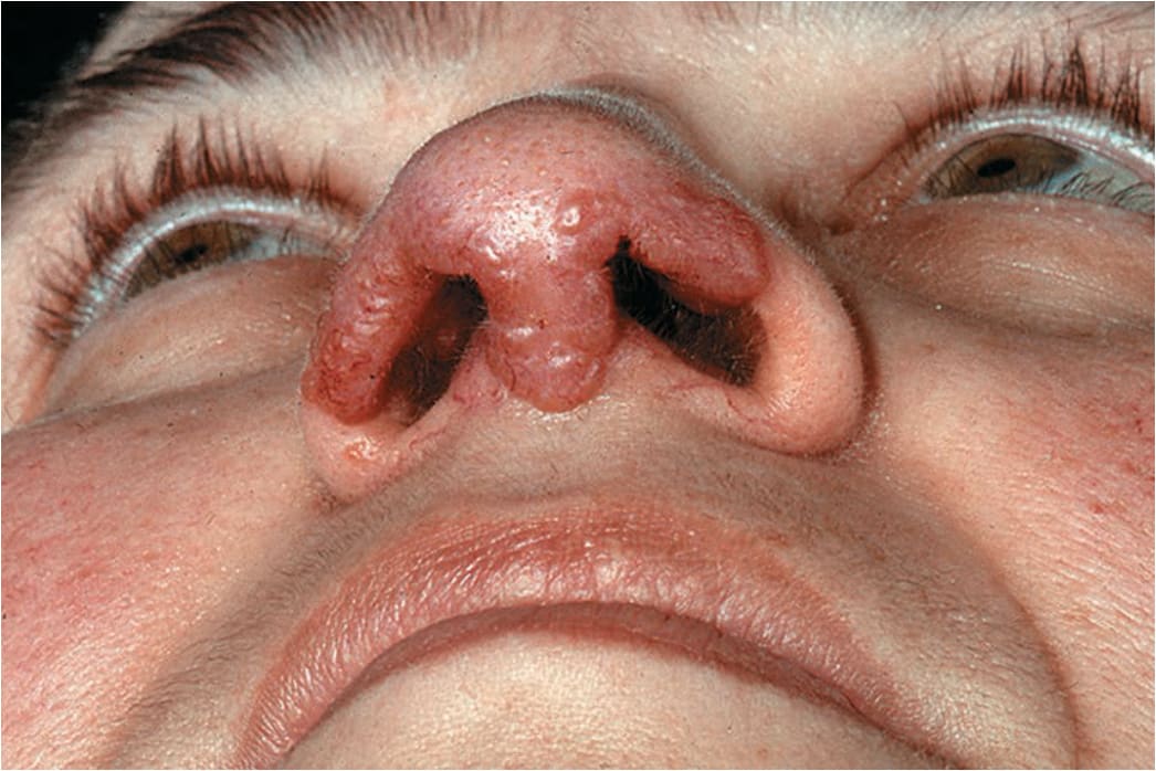
FIGURE 14-66 • Sarcoidosis Brownish-to-purple papules coalescing to irregular plaques, occurring on the nose of this woman who also had massive pulmonary involvement. Blanching with a glass slide reveals “apple-jelly” color in the lesions.
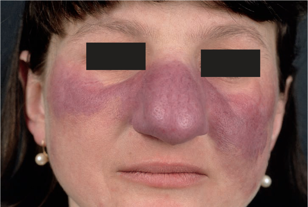
FIGURE 14-67 • Sarcoidosis This is the classic appearance of “lupus pernio” with violaceous, soft, doughy infiltrations on cheeks and nose, which is grossly enlarged.
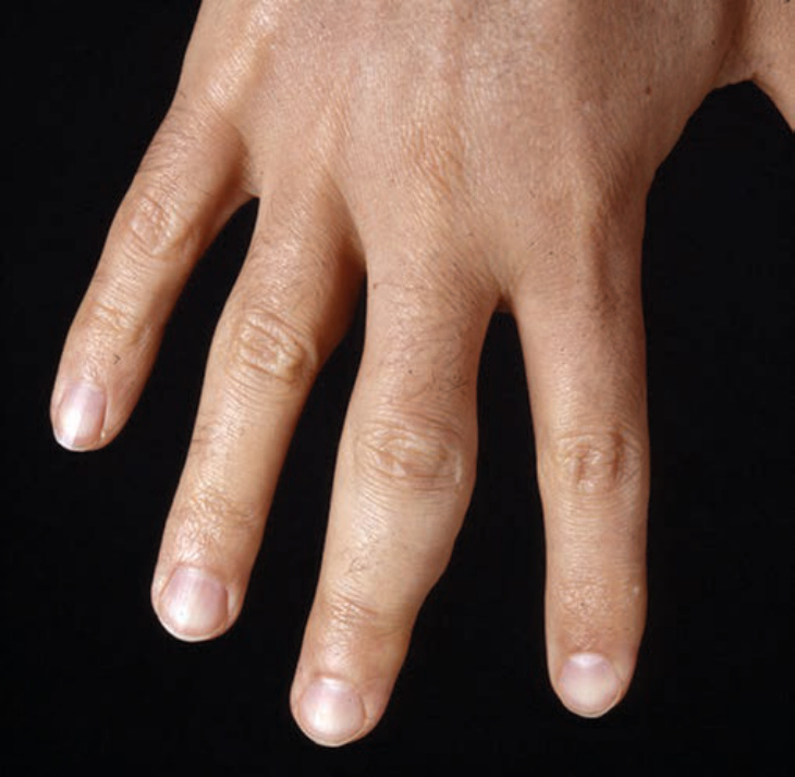
FIGURE 14-68 • Sarcoidosis Firm swelling of the third digit resulting from osteitis cystica in a 52-year-old man with pulmonary involvement.
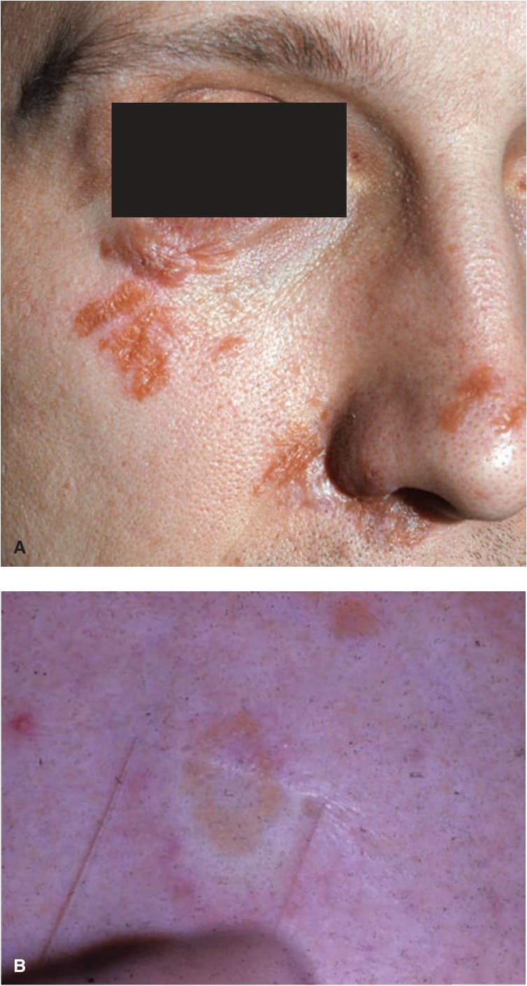
FIGURE 14-69 • (A) Sarcoidosis in scars Bizarre scars are almost replaced by brownish-red sarcoidal infiltrates. Years previously this man had a motorcycle accident suffering facial lesions when he skidded on a dirt road. (B) Diascopy of sarcoidal lesions. Note the change in color of these infiltrative papules under pressure from a glass slide. The so-called “apple jelly” discoloration is seen. (Used with permission of Dr. Kenneth Greer.)