NECROBIOSIS LIPOIDICA
NECROBIOSIS LIPOIDICA ICD-10: E14.640
• Necrobiosis lipoidica (NL) is a cutaneous disorder often, but not always, associated with diabetes mellitus.
• Young adults, early middle age, but not uncommon in juvenile diabetics. Female to male ratio: 3:1.
• Incidence: From 0.3% to 3% of diabetic individuals. One-third of patients have clinical diabetes, one-third have abnormal glucose tolerance only, and one-third have normal glucose tolerance.
• The severity of NL is not related to the severity of diabetes. Control of the diabetes has no effect on the course of NL.
• Lesion starts as brownish-red or skin-colored papule that slowly evolves into a well-demarcated waxy plaque of variable size (Fig. 15-4). The sharply defined and slightly elevated border retains a brownish-red color, whereas the center becomes depressed and acquires a yellow-orange hue. Multiple telangiectasias of variable size are noted. Larger lesions formed by centrifugal enlargement or merging of smaller lesions acquiring a serpiginous or polycyclic configuration. Ulceration may occur and healed ulcers result in depressed scars. Burned-out lesions are tan with telangiectasia.
• Usually one to three lesions; >80% occur on the shin; at times symmetric. Less commonly on feet, arms, trunk, or face and scalp; rarely may be generalized.
• Dermatopathology: Sclerosis, obliteration of the bundle pattern of collagen → necrobiosis, surrounded by concomitant granulomatous infiltration in lower dermis. Microangiopathy.
• Biopsy confirmation is usually not necessary; however, biopsy may be required in early stages to rule out granuloma annulare (which frequently coexists with NL), sarcoidosis, or xanthoma.
• Glucocorticoids. Topical: Under occlusion is helpful; however, ulcerations may occur when NL is occluded. Intralesional: triamcinolone, 5 mg/mL, into active lesions or lesion margins usually arrests extension of plaques of NL. Ulceration: Most ulcerations within NL lesions heal with local wound care; if not, excision of the entire lesion with grafting may be required.
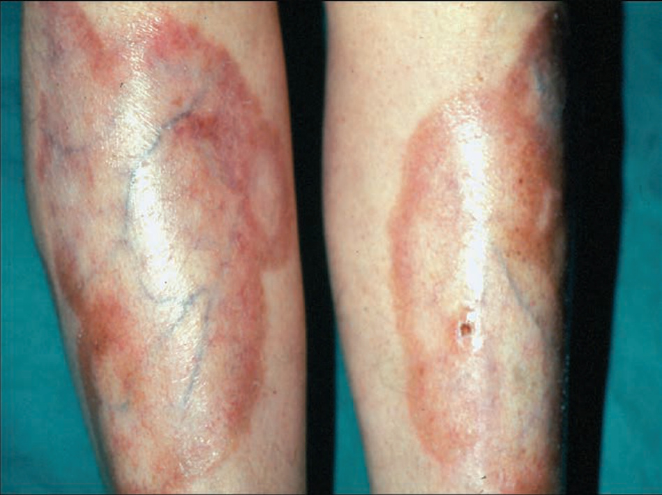
FIGURE 15-4 • Necrobiosis lipoidica diabeticorum A large, symmetric plaque with active tan-pink, yellow, well-demarcated, raised, firm border and a yellow center in the pretibial region of a 28-year-old diabetic female. The central parts of the lesion are depressed with atrophic changes of epidermal thinning and telangiectasia against a yellow background. (Used with permission of Dr. Kenneth Greer.)