TUBEROUS SCLEROSIS (TS)
TUBEROUS SCLEROSIS (TS) ICD-10: Q85.1
• Tuberous sclerosis is an autosomal-dominant disease arising from a genetically programmed hyperplasia of ectodermal and mesodermal cells and manifested by a variety of lesions in the skin, CNS (hamartomas), heart, kidney, and other organs.
• The principal early manifestations are the triad of seizures, mental retardation, and congenital white spots.
• Facial angiofibromata are pathognomonic but do not appear until the 3rd or 4th year.
EPIDEMIOLOGY
INCIDENCE In mental institutions, 1:100 to 1:300; in general population, 1:20,000 to 1:100,000. AGE OF ONSET Infancy. SEX Equal incidence. RACE All races. HEREDITY Autosomal dominant. TS is caused by mutations in a tumor-suppressor gene, either TSCS1 or TSCS2. TSCS1 maps to chromosome 9q34. TSCS2 maps to 16p13.3.
CLINICAL MANIFESTATION
White macules are present at birth or appear in infancy (>80% occur by 1 year of age, 100% appear by 2 years); >20% of angiofibromata are present at 1 year of age, 50% occur by 3
years. Seizures (infantile spasms) occur in 86%; the earlier the onset of seizures, the worse the mental retardation. Mental retardation (49%). SKIN LESIONS (96% incidence).
Hypomelanotic Macules “Off-white”; one or many, usually more than three. Polygonal or “thumbprint,” 0.5 to 2 cm; lance ovate or “ashleaf” spots (Fig. 16-2), 3 to 4 cm (up to 12 cm); tiny white “confetti” macules, 1 to 2 mm (Fig. 16-3). White macules occur on trunk (>), lower extremities (>), upper extremities (7%), head and neck (5%). White macules shine up with Wood light (Fig. 16-2B).
Angiofibromas 0.1 to 0.5 cm, dome-shaped and smooth, exhibiting red or skin color (Fig. 16-4). Occur in the center of the face.
A B
A B
They are firm and disseminated but may coalesce; termed adenoma sebaceum but represent angiofibromas (present in 70%).
Plaques Represent connective tissue nevi (“shagreen” patch), present in 40%; skin colored; occur on the back and buttocks (Fig. 16-5B).
Periungual Papules or Nodules Ungual fibromas (Koenen tumors) present in 22%, arise late in childhood, and have the same pathology (angiofibroma) as facial papules (Fig. 16-5A).
ASSOCIATED SYSTEMS
CNS (tumors producing seizures), eye (gray or yellow retinal plaques, 50%), heart (benign rhabdomyomas), and hamartomas of mixed cell type (kidney, liver, thyroid, testes, and GI system).
LABORATORY EXAMINATIONS
DERMATOPATHOLOGY White Macules Decreased number of melanocytes, decreased melanosome size, decreased melanin in melanocytes and keratinocytes.
Angiofibromata Proliferation of fibroblasts, increased collagen, angioneogenesis, capillary dilatation, and absence of elastic tissue. BRAIN PATHOLOGY “Tubers” are gliomas. IMAGING Skull X-Ray Multiple calcific densities.
CT Scan Ventricular deformity and tumor deposits along the striothalamic borders.
MRI Subependymal nodules.
Electroencephalography Abnormal.
Renal Ultrasound Reveals renal hamartoma.
DIAGNOSIS
More than five ash leaf macules (Fig. 16-2) in an infant are highly suggestive. Confetti spots (Fig. 16-2) are virtually pathognomonic. Evaluate the patient with a study of the family members and by obtaining various types of imaging as well as electroencephalography. Mental retardation and seizures may be absent.
DIFFERENTIAL DIAGNOSIS
WHITE SPOTS Focal vitiligo, nevus anemicus, tinea versicolor, nevus depigmentosus, pityriasis alba, and postinflammatory hypomelanosis. ANGIOFIBROMAS Tricholemmona, syringoma, skin-colored papules on the face, and dermal nevi. Note: Angiofibromata of the face (Fig. 16-4) have been mistaken for and treated as acne vulgaris or rosacea. PERIUNGUAL FIBROMAS Verruca vulgaris.
COURSE AND PROGNOSIS
Tuberous sclerosis is a serious autosomal disorder that causes major problems in behavior, caused by mental retardation, and in therapy, the aim is to control the seizures. In severe cases, 30% die before the fifth year of life, and 50 to 75% die before reaching adult age. Malignant gliomas are not uncommon. Genetic counseling is imperative.
MANAGEMENT
PREVENTION Counseling. TREATMENT Laser surgery for angiofibromas. Support Organization: https://www. tscalliance.org
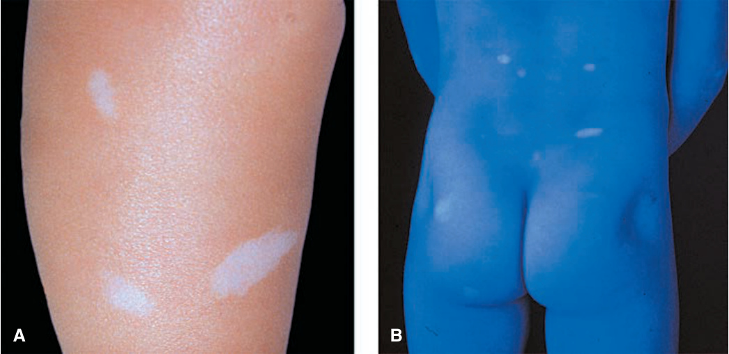
FIGURE 16-2 • Tuberous sclerosis: ash-leaflet hypopigmented macules (A) Three well-demarcated, elongated (ash-leaflet shaped), hypomelanotic macules on the lower leg of a child with tan skin. (B) Ash-leaflet hypomelanotic macules in pale skin are better visualized under Wood light where they light up.
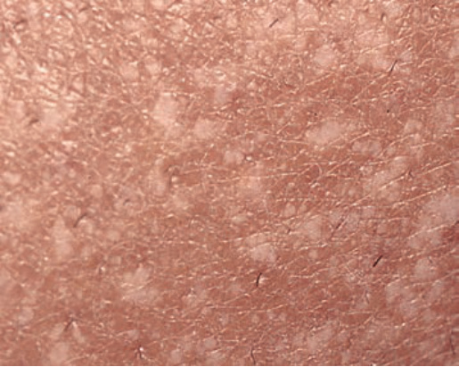
FIGURE 16-3 • Tuberous sclerosis: “confetti” macules Multiple, discrete, small, confetti-like, hypopigmented macules of variable size on the leg. These lesions are pathognomonic.
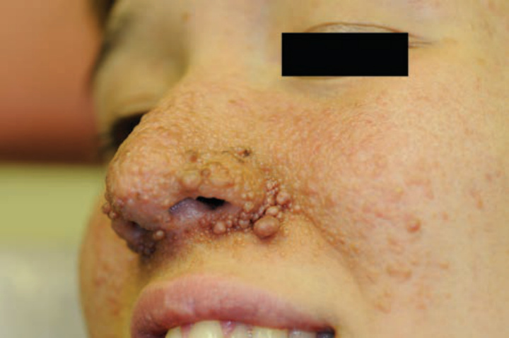
FIGURE 16-4 • Tuberous sclerosis: angiofibromas Confluent, small, angiomatous (erythematous, glistening) papules on the cheek and nose. These lesions were not present during the first few years of life; appeared only after the first 5 years of life. (Used with permission of Dr. Barrett Zlotoff.)
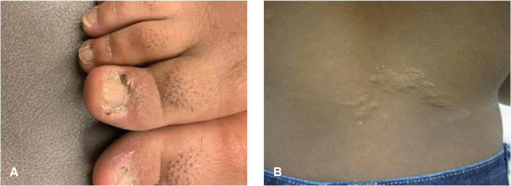
FIGURE 16-5 • Tuberous sclerosis (A) Periungual fibroma (Koenen tumor). (B) Shagreen patch, slightly elevated, skin colored. This represents a connective tissue nevus. (Used with permission of Dr. Jennifer Tan.)