PRESSURE ULCERS
PRESSURE ULCERS ICD-10: L89
• Pressure ulcers develop at body-support interfaces over bony prominences as a result of external compression of the skin, shear forces, and friction, which produce ischemic tissue necrosis.
• Occurs in patients who are obtunded mentally or have diminished sensation (as in spinal cord disease) in the affected region. Secondary infection results in localized cellulitis, which can extend locally into bone or muscle or into the bloodstream.
EPIDEMIOLOGY
AGE OF ONSET Any age, but the greatest prevalence of pressure ulcers is in elderly, chronically bedridden patients. SEX Equally prevalent in both sexes.
PATHOGENESIS
Risk factors: Inadequate nursing care, diminished sensation/immobility (obtunded mental status, spinal cord disease), hypotension, fecal or urinary incontinence, the presence of fracture, hypoalbuminemia, and poor nutritional status. Infection also impairs or prevents healing.
CLINICAL MANIFESTATION
SKIN LESIONS Clinical Categories of Pressure Ulcers Early change: Localized erythema that blanches on pressure. Stage I: Nonblanching erythema of intact skin. Stage II: Necrosis, superficial or partial thickness involving the epidermis and/or dermis. Bullae → necrosis of dermis (black) → shallow ulcer. Stage III: Deep necrosis, crateriform ulceration with full-thickness skin loss (Fig. 17-17); damage or necrosis can extend down to, but not through, fascia. Stage IV: Full-thickness necrosis (→ ulcer ation) with involvement of supporting
structures such as muscle and bone (Fig. 17-18). May enlarge to many centimeters. May or may not be tender. Borders of ulcers may be undetermined. Well-established pressure ulcers with devitalized tissue at the base (eschar) have a higher chance of secondary infection.
Distribution Occur over bony prominences: sacrum (60%) > ischial tuberosities, greater trochanter, heel (Fig. 17-18) > elbow, knee, ankle, and occiput.
LABORATORY EXAMINATIONS Hematologic Studies
WOUND CULTURE For aerobic and anaerobic bacteria. BLOOD CULTURE Bacteremia often follows manipulation of ulcer. PATHOLOGY Skin Biopsy Epidermal necrosis with eccrine duct and gland necrosis. Deep ulcers show wedge-shaped infarcts of the subcutaneous tissue.
Bone Biopsy Essential for diagnosing continuous osteomyelitis.
DIAGNOSIS AND DIFFERENTIAL DIAGNOSIS
Usually made clinically. Differential diagnosis includes infectious ulcer (actinomycotic infection, deep fungal infection, chronic
herpetic ulcer), thermal burn, malignant ulcer, pyoderma gangrenosum, and rectocutaneous fistula.
COURSE AND PROGNOSIS
If pressure is relieved, some changes are reversible. Osteomyelitis occurs in nonhealing pressure ulcers (32% to 81%). Septicemia is associated with a high mortality rate. With proper treatment, stages I and II ulcers heal in 1 to 4 weeks and stages III and IV ulcers heal in 6 to >12 weeks.
MANAGEMENT
PROPHYLAXIS IN AT-RISK PATIENTS Reposition patient every 2 hours (more often if possible); inspect for areas of skin breakdown over pressure points; minimize friction and shear forces.
• Use interface air mattress to reduce compression.
• Minimize skin exposure to excessive moisture from incontinence, perspiration, or wound drainage.
• Evaluate and correct nutritional status; consider supplements of vitamin C and zinc.
STAGES I AND II ULCERS Topical antibiotics (not neomycin) under moist sterile gauze may be sufficient for early erosions. Hydrogels, alginate or hydrocolloid dressings. STAGES III AND IV ULCERS Surgical management: Debridement of necrotic tissue, bony prominence removal, flaps, and skin grafts. Vacuum-assisted closure (VAC). Recombinant platelet-derived growth factors. Some data with hyperbaric oxygen, electrical stimulation, and warm-up therapy. INFECTIOUS COMPLICATIONS Prolonged course of antimicrobial agent depending on sensitivities and with surgical debridement of necrotic bone in osteomyelitis.
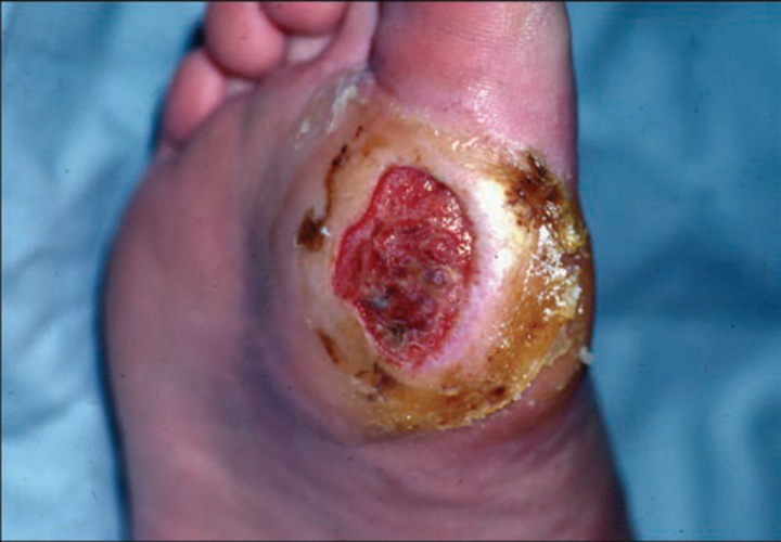
FIGURE 17-17 • Pressure ulcer, stage III Well-demarcated crateriform ulcer with full-thickness skin loss at the base of the toe. (Used with permission of Dr. Kenneth Greer.)
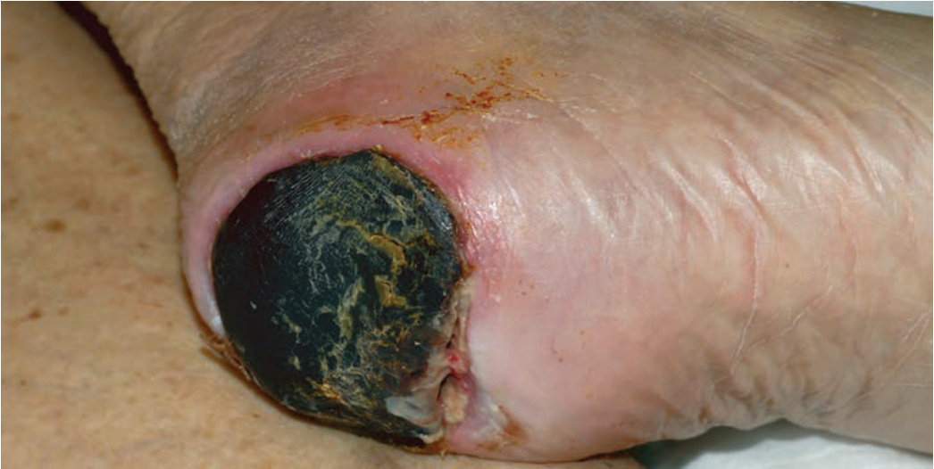
FIGURE 17-18 • Pressure ulcer, stage IV on the heel The black necrosis seen here extended into the calcaneal bone which also had to be debrided.