GLUCAGONOMA SYNDROME
GLUCAGONOMA SYNDROME ICD-10: M8152/0
• Glucagonoma syndrome is a rare but well-described clinical entity caused by excessive production of glucagon in an α-cell tumor of the pancreas.
• Characterized by superficial migratory necrolytic erythema (MNE) with erosions that crust and heal with hyperpigmentation.
• Inflammatory patches and red plaques (Figs. 19-14 and 19-15) of gyrate, circinate, arcuate, or annular shape that enlarge with central clearing, resulting in geographic areas that become confluent (Fig. 19-15). Borders show vesiculation to bulla formation, crusting, and scaling.
• Lesions involve perioral and perigenital regions and flexures and intertriginal areas.
• Fingertips are red, shining, and erosive (Fig. 19-16).
• There is glossitis, angular cheilitis (Fig. 19-14), and blepharitis.
• General examination reveals wasting and malnutrition.
• Most cases are associated with glucagonoma, but the pathogenesis of MNE is not known. There exists MNE without glucagonoma.
• Differential diagnosis: Includes all moist red plaque(s): Acrodermatitis enteropathica, zinc deficiency, pustular psoriasis, mucocutaneous candidiasis, Hailey–Hailey disease (familial pemphigus).
• Laboratory: Fasting plasma glucagon level increased to >1,000 ng/L (normal 50 to 250 ng/L) and makes the diagnosis. There is also hyperglycemia, reduced glucose tolerance, severe malabsorption, gross hypoaminoacidemia, and low serum zinc. CT scan angiography will locate tumors within pancreas and metastases in the liver. MRI or PET scanning may be used if CT scans are unrevealing.
• Dermatopathology of early skin lesions shows band-like upper epidermal necrosis with retention of pyknotic nuclei and pale keratinocyte cytoplasm.
• Prognosis depends on the aggressiveness of the glucagonoma. Hepatic metastases have occurred in 75% of patients at the time of diagnosis. If these are slow growing, patients may have prolonged survival, even with metastatic disease.
• MNE responds poorly to all types of therapy. Some cases have responded partially to zinc replacement, hydration and IV administration of amino or fatty acids. MNE resolves after tumor excision. However, surgical excision of glucagonoma achieves a cure in only 30% of cases because of persistent metastases (usually liver). Such patients can be treated with chemotherapy (streptozocin and doxorubicin) and newer agents under investigations (everolimus and sunitinib). Octreotide can be used to suppress glucagon production.
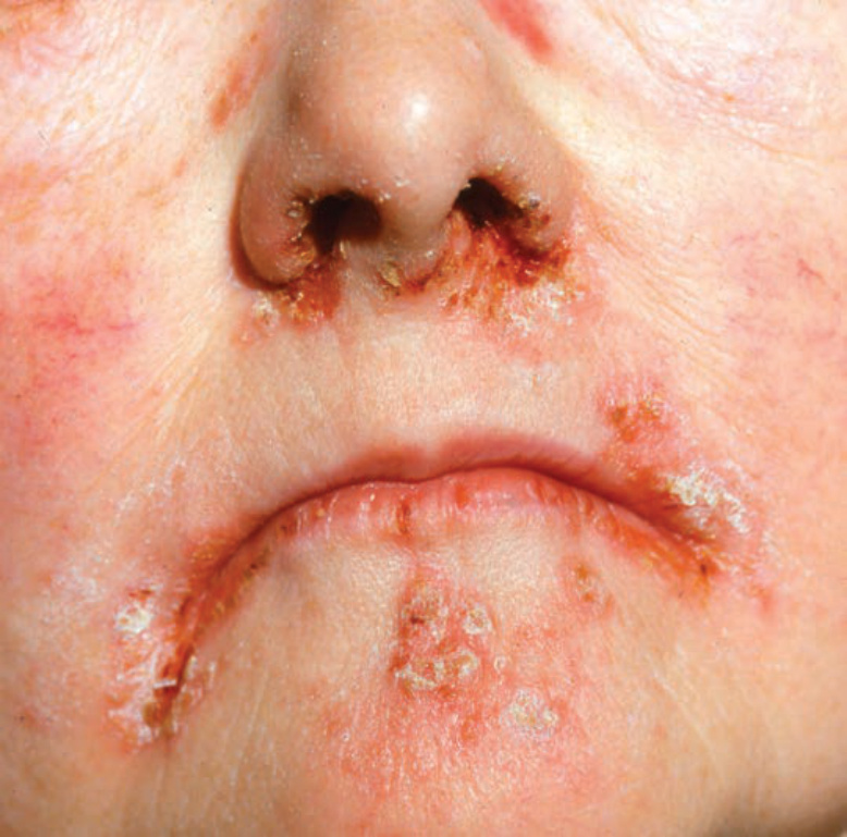
FIGURE 19-14 • Glucagonoma syndrome: migratory necrolytic erythema Inflammatory dermatosis with angular cheilitis, inflammatory, scaly, erosive, and crusted plaques and fissures around the nose and mouth.
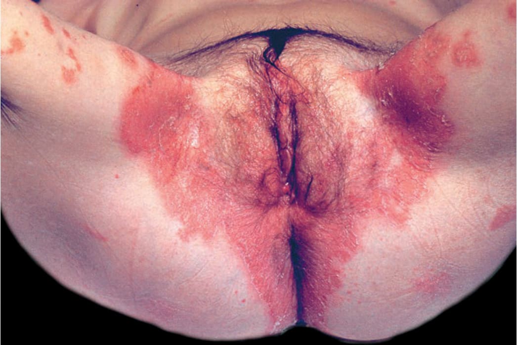
FIGURE 19-15 • Glucagonoma syndrome: migratory necrolytic erythema Polycyclic erosions in the anogenital gluteal and sacral regions. Sharply defined with necrotic flaccid epidermis still covering part of these erosions.
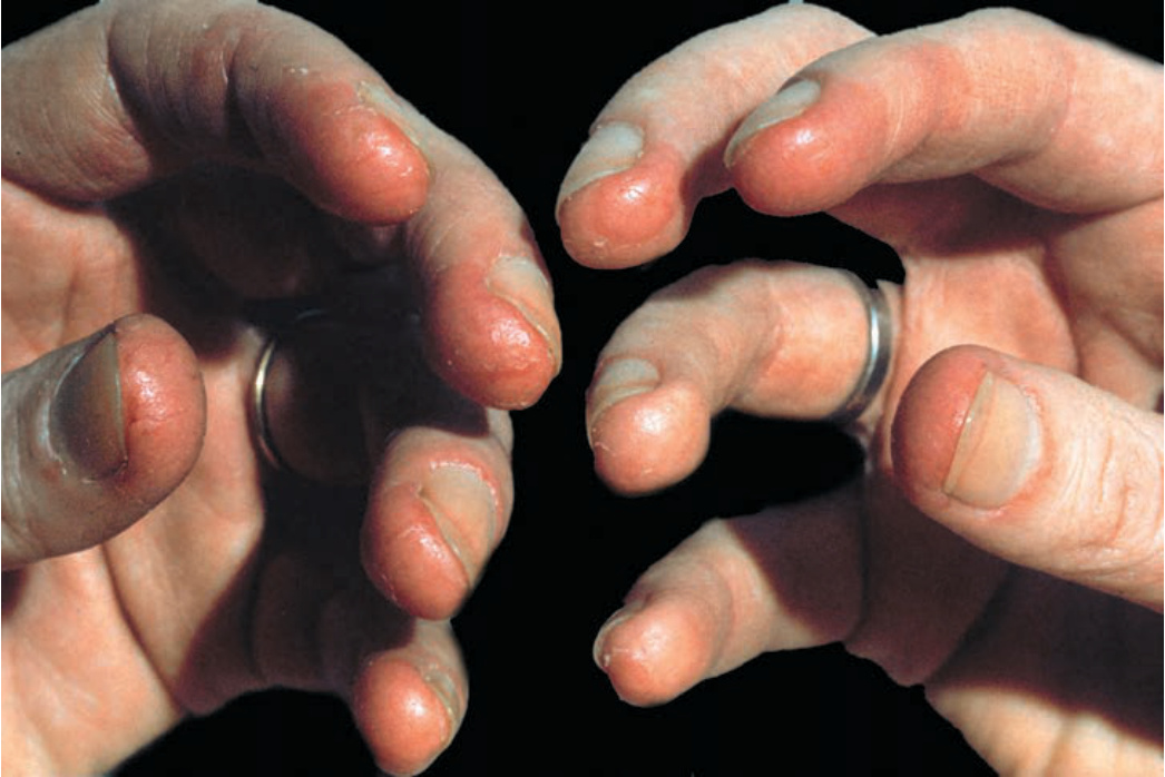
FIGURE 19-16 • Glucagonoma syndrome Fingertips are red, glistening, and partially erosive.