DERMATOFIBROSARCOMA PROTUBERANS (DFSP)
DERMATOFIBROSARCOMA PROTUBERANS (DFSP) ICD10: C44.9
• Tissue sarcoma arising in the dermis and often penetrates into fat.
• Etiology is unknown but has been reported to arise in scars. It is caused by a translocation between chromosome 17 and 22 creating the COL1A1-PDGFB fusion gene.
• Incidence is low, approximately 1 to 5 people per million.
• There appears to be a higher rate in African Americans compared to Caucasians; females are overrepresented. Tumor growth may be accelerated during pregnancy.
• Peak age of incidence is between 20 and 50 years of age.
• The tumor may present as a nonspecific plaque often resembling a keloid, with purplish to brownish coloration (Fig. 21-22). Other clinical variants include the Bednar tumor (pigmented), giant cell fibroblastoma (usually in children), and fibrosarcomatous DFSP which carries the highest risk of metastasis.
• Diagnosis is by histopathology, which shows a spindle cell proliferation often arranged as whorls (so-called herringbone pattern). Clinical distinction from dermatofibroma can be made by showing CD34 staining. Of note, fibrosarcomatous areas in DFSP may fail to stain with CD34.
• Treatment is usually surgical. Mohs micrographic surgery has been used. Due to the molecular basis for this tumor, Imatinib has shown promise, mostly to attempt decrease in tumor size prior to surgical intervention. Around 50% response rates are seen. Radiation is also an option. Standard chemotherapy is ineffective.
• If cleared by resection, prognosis is good.
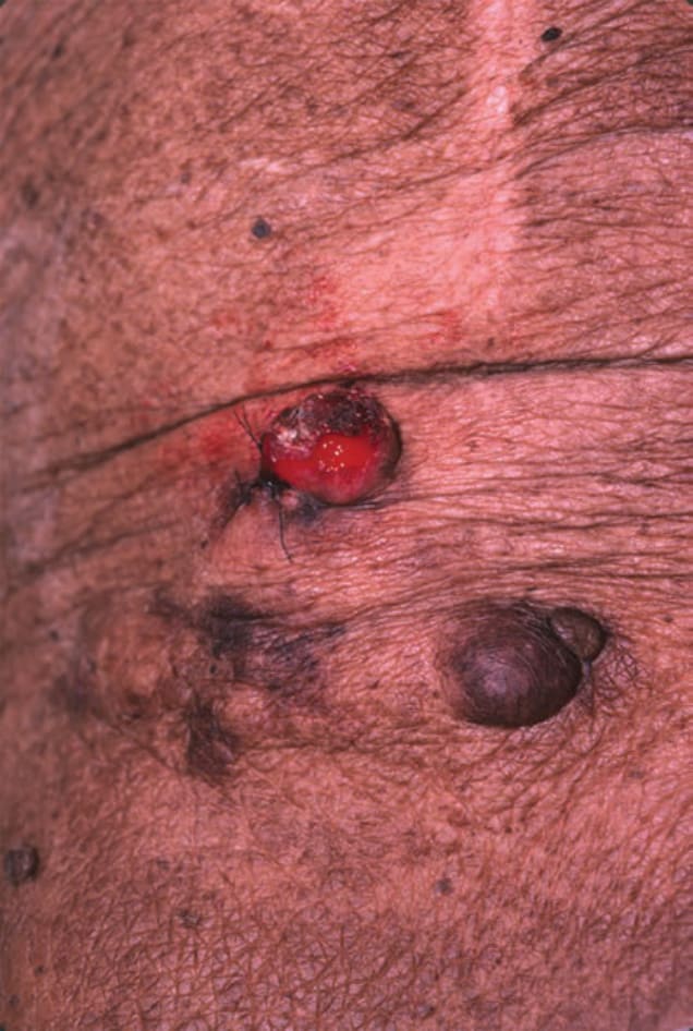
FIGURE 21-22 • Dermatofibrosarcoma protuberans Tumors may present as nodules coalescing into plaques, often resembling a keloid. Though the tumor itself was slow-growing, peripheral infiltration into skin differentiates this lesion from a hypertrophic scar or keloid. (Used with permission of Dr. Kenneth Greer.)