EXANTHEMATOUS DRUG REACTIONS
EXANTHEMATOUS DRUG REACTIONS ICD-10: T88.7
• An exanthematous drug reaction (EDR) (eruption) is an adverse hypersensitivity reaction to an ingested or parenterally administered drug that mimics a measles-like viral exanthem.
• Most common type of cutaneous drug reaction.
• Systemic involvement is low.
• Drugs with a high probability of reaction (3% to 5%): Penicillin and related antibiotics, carbamazepine, allopurinol, and gold salts (10% to 20%). Medium probability: Sulfonamides (bacteriostatic, antidiabetic, diuretic), nonsteroidal anti-inflammatory drugs (NSAIDs), hydantoin derivatives, isoniazid, chloramphenicol, erythromycin, and streptomycin. Low probability (<1%): Barbiturates, benzodiazepines, phenothiazines, and tetracyclines.
• Prior Drug Sensitization. Patients with a prior history of exanthematous drug eruption will most likely develop a similar reaction if rechallenged with the same drug.
• Sensitization occurs during administration or after completing the course of drugs; peak incidence is usually at ninth day after administration. However, EDR may occur at any time between the first day and 3 weeks after the beginning of treatment. Reaction to penicillin can begin ≥2 weeks after drug is discontinued. In previously sensitized patient, eruption starts within 2 or 3 days after readministration of drug.
• Usually quite pruritic. Painful skin lesions suggest development of a more serious ACDR, such as toxic epidermal necrolysis (TEN).
• Systems Review. ± Fever and chills.
• Skin Lesions. Macules and/or papules, a few millimeters to 1 cm in size (Fig. 23-1). Bright or “drug” red. In time, lesions become confluent forming large macules, polycyclic/gyrate erythema, reticular eruptions, sheet-like erythema (Fig. 23-1), or erythroderma; also erythema multiforme-like. Purpura may be seen in lesions of the legs. In individuals with thrombocytopenia, exanthematous eruptions can mimic vasculitis because of intralesional hemorrhage. Scaling and/or desquamation may occur with healing.
• Distribution. Symmetric (Fig. 23-1). Almost always occurs on the trunk and extremities. In children, it may be limited to the face and extremities. Commonly EDR affect upper trunk and upper extremities before affecting the lower trunk and lower extremities.
• Mucous Membranes. Enanthem on buccal mucosa.
• Laboratory. Peripheral eosinophilia. Dermatopathology: Perivascular lymphocytes and eosinophils.
• Differential diagnosis includes all exanthematous eruptions: Viral exanthem, secondary syphilis, atypical pityriasis rosea, and early widespread allergic contact dermatitis.
• After discontinuation of the drug, the rash usually fades over 10–20 days with exfoliation, similar to a sunburn. However, it may worsen for a few days. The eruption may also begin after the drug has been discontinued. Eruption usually recurs with rechallenge.
• The definitive step in management is to identify the offending drug and discontinue it. Oral antihistamine can alleviate pruritus. Glucocorticoids. Potent Topical Preparation, Oral or IV. If the offending drug cannot be substituted or omitted, systemic glucocorticoids can be administered to treat the ACDR. Prevention. Patients must be aware of their specific drug hypersensitivity and that other drugs of the same class can cross-react. Wearing a medical alert bracelet is advised.
REACTIONS TO SPECIFIC DRUGS (SELECTED)
Abacavir HLA-B*5701 and HHV-6 are risk factors. Can lead to cardiovascular collapse. HLA testing is essential.
Allopurinol Incidence: 5%. Begins on the face, spreads rapidly to all areas; may occur in photodistribution. Onset: 2 to 3 weeks after initiation of therapy. Associated findings: Facial edema; systemic vasculitis, especially involving kidneys. The rash may fade in spite of continued administration. Higher doses, kidney disease and HLA-B*5801 are risk factors. Avoid thiazides use concurrently.
Ampicillin, Amoxicillin In up to 100% of patients with EBV or CMV mononucleosis syndrome. Increased incidence of EDR to penicillins in patients taking allopurinol. Ten percent cross-react with cephalosporins.
Carbamazepine Morphology: diffuse erythema; severe erythroderma may follow. Site: Begins on the face, then spreads rapidly to all areas; may occur in photodistribution. Onset: 2 weeks after initiation of therapy. Associated findings: Facial edema. HLA-B1502 and HLA3101 are risk factors.
Hydantoin Derivatives Macular → confluent erythema. Begins on the face, then spreads to trunk and extremities. Onset: 2 weeks after initiation of therapy. Associated findings: Fever, peripheral eosinophilia; facial edema; lymphadenopathy (can mimic lymphoma histologically).
Lamotrigine Younger population at risk and those with concurrent valproic acid. Follow dose escalation protocol.
Nevirapine Females are at greatest risk, those with HLA-B*3505 and those who have not complied to lead-in dosing. Risk of progression to toxic epidermal necrolysis. Sulfonamides Occurs in up to 50% to 60% of HIV/AIDS-infected patients (trimethoprim sulfamethoxazole). Patients sensitized to one sulfa-based drug may cross-react with another sulfa drug in 20%.
Warfarin Female sex, obesity, and hereditary protein C and S deficiency are risk factors. Avoid large doses initially. Drug-induced urticaria/angioedema usually resolves within hours, days, or weeks after the causative drug is withdrawn.
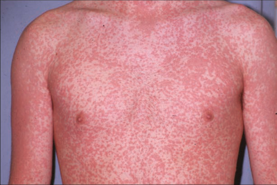
FIGURE 23-1 • Exanthematous drug eruption Symmetrically arranged, brightly erythematous macules and papules, discrete in some areas, and confluent in others, on the trunk and the extremities. (Used with permission of Dr. Kenneth Greer.)