INTERTRIGO
INTERTRIGO ICD-10: L30.4
• Intertrigo (Latin inter, “between”; trigo, “rubbing”).
• Inflammation of opposed skin (inframammary regions, axillae, groins, gluteal folds, and redundant skin folds of obese persons). May represent inflammatory dermatosis or superficial colonization or infection.
• Dermatoses occurring in intertriginous skin include intertriginous psoriasis, seborrheic dermatitis, Hailey–Hailey disease, and Langerhans cell histiocytosis. S. aureus and streptococcus can cause secondary infection of these dermatoses.
INFECTIOUS INTERTRIGO
BACTERIAL
• Beta-hemolytic streptococci. Group A (Fig. 25-5), group B, and group G (Fig. 25-6). Streptococcal intertrigo can progress to soft-tissue infection (Fig. 25-6).
• S. aureus. Often gains entry into the skin via hair follicles, causing folliculitis and furuncles.
• Pseudomonas aeruginosa (Fig. 25-7A involving skin 25-7B involving nail).
• C. minutissimum (erythrasma) (Figs. 25-1 and 25-2).
• K. sedentarius (pitted keratolysis) (Fig. 25-3).
CLINICAL MANIFESTATION
Usually asymptomatic. Discomfort usually indicates infection rather than colonization. Soft-tissue infection can gain entry in S. aureus or streptococcal intertrigo.
DIAGNOSIS
Identify pathogen by bacterial culture, Wood’s lamp examination, or KOH preparation.
TREATMENT
Identify and treat pathogen.
A
B
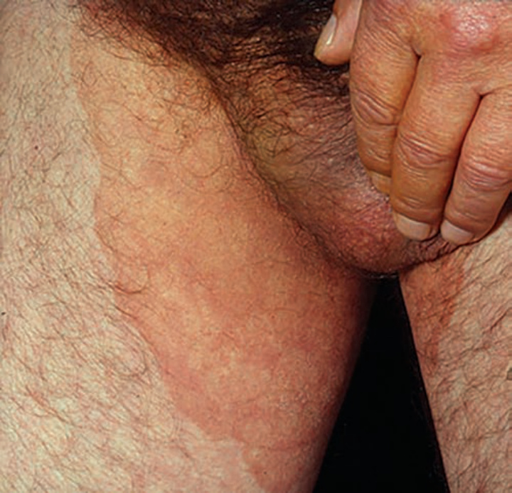
FIGURE 25-1 • Erythrasma: Groins Sharply marginated, tan patches in the genitocrural fold. Wood lamps demonstrates bright coral-red fluorescence differentiating erythrasma from intertriginous psoriasis. KOH preparation was negative for hyphae.
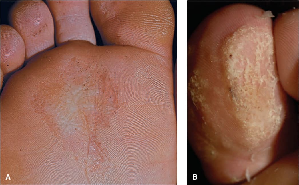
FIGURE 25-3 • A, B Pitted keratolysis: Plantar The stratum corneum of the plantar skin (A) and toe-pad (B) shows confluent multiple, confluent “pits” (defects in the stratum corneum).
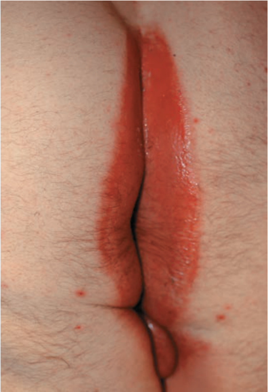
FIGURE 25-5 • Intergluteal intertrigo: Group A streptococcus A painful moist erythematous plaque in a male with intertriginous psoriasis, with foul odor. Infection resolved with penicillin VK.
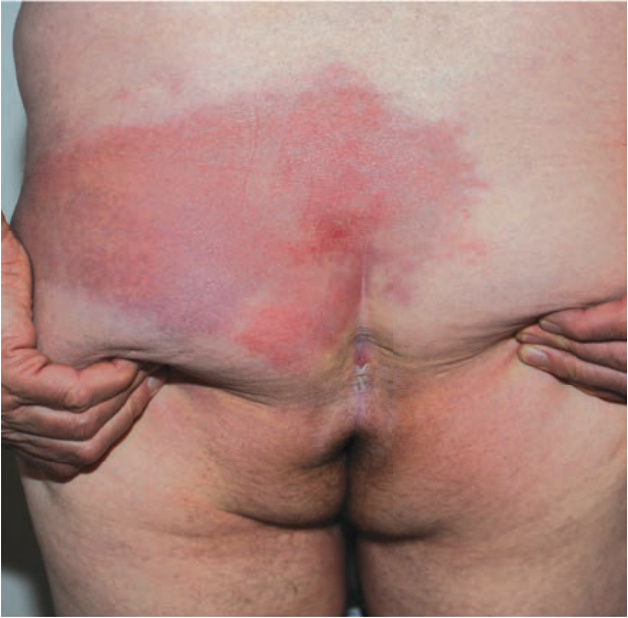
FIGURE 25-6 • Erysipelas: Group G streptococcus A 65-year-old male with sharply marginated erythematous plaque on buttocks. Portal of entry of infection was intergluteal intertrigo.
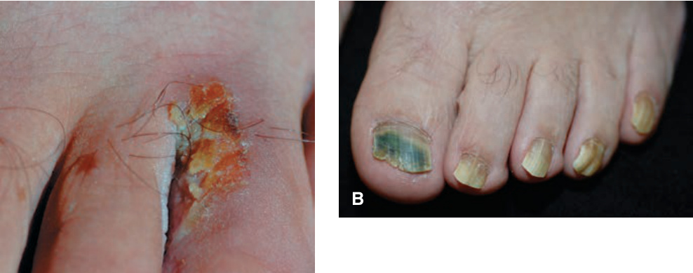
FIGURE 25-7 • A, B Webspace intertrigo and Pseudomonal toenail infection: P. aeruginosa Erosion of a webspace of the foot with a bright red base and surrounding erythema. Tinea pedis (interdigital and moccasin patterns) and hyperhidrosis were also present, which facilitated growth of Pseudomonas. (B) Shows Pseudomonas infection of the nail plate leading to a green-brown discoloration.