IMPETIGO
IMPETIGO ICD-10: B08.0
• Etiology. S. aureus; GAS.
• Portal of Entry. Impetigo occurs adjacent to the site of S. aureus colonization such as the nares. Secondary infection of (1) minor breaks in the epidermis (impetiginization), (2) preexisting dermatoses, (3) other infections such as eczema herpeticum, or (4) wounds.
• Clinical Manifestation. Honey-colored crusted erosions.
• Treatment
• Reduced colonization.
• Topical antibiotic to infected and colonized sites; systemic antibiotic.
EPIDEMIOLOGY AND ETIOLOGY
• S. aureus: Methicillin-sensitive (MSSA) and methicillin-resistant (MRSA). Bullous impetigo: Local production of epidermolytic toxin A–producing S. aureus, which also causes staphylococcal scalded skin syndrome.
• Beta-hemolytic streptococcus: Group A.
S. aureus and GAS are not members of human skin microbiome. They may transiently colonize skin and cause superficial infections. DEMOGRAPHY Secondary infections, any age. Primary infections most often occur in children.
PORTALS OF ENTRY OF INFECTION Minor breaks in the skin most commonly. Facial lesions usually associated with S. aureus colonization of nares. Dermatoses such as atopic dermatitis or Hailey–Hailey disease. Traumatic wounds. Bacterial infections occur in other cutaneous infections.
CLINICAL MANIFESTATION
Superficial infections often asymptomatic. Ecthyma may be painful and tender. IMPETIGO Erosions with crusts (Figs. 25-8 and 25-9). Golden-yellow crusts are often seen in impetigo but are hardly pathognomonic; 1- to >3-cm lesions; central healing often apparent
if lesions present for several weeks (Fig. 25-9). Arrangement: Scattered, discrete lesions; without therapy, lesions may become confluent; satellite lesions occur by autoinoculation. Secondary infection of various dermatoses is common (Figs. 25-10 and 25-11).
BULLOUS IMPETIGO Superficial blisters containing clear yellow or slightly turbid fluid with erythematous halo, arising on normal-appearing skin. Bullous lesions rupture easily, revealing shallow moist erosions (Figs. 25-12 and 25-13). Distribution: More common in intertriginous sites.
ECTHYMA Ulceration with a thick adherent crust (Fig. 25-14). Lesions may be tender, indurated. Usually occurs at occluded sites (common in homeless or soldiers in trenches during combat who do not or cannot change boots).
DIFFERENTIAL DIAGNOSIS
IMPETIGO Excoriation, contact dermatitis, herpes simplex, epidermal dermatophytosis, and scabies. INTACT BULLAE Acute contact dermatitis, insect bites, thermal burns, and porphyria cutanea tarda (PCT) (dorsa of hands). ECTHYMA Excoriations, insect bites.
DIAGNOSIS
Clinical findings confirmed by culture: S. aureus, commonly; failure of oral antibiotic suggests MRSA. GAS.
COURSE
Untreated, lesions of impetigo become more extensive and develop into ecthyma.
With adequate treatment, prompt resolution. Lesions can progress to deeper skin and soft-tissue infections. Nonsuppurative complications of GAS infection include guttate psoriasis, scarlet fever, and glomerulonephritis. Ecthyma may heal with scarring. Recurrent S. aureus or GAS infections can occur because of the failure to eradicate pathogen or by recolonization. Undiagnosed MRSA infection does not respond to usual oral antibiotics given for methicillin-sensitive S. aureus.
TREATMENT
PREVENTION Benzoyl peroxide wash. Check family members for signs of impetigo. Ethanol or isopropyl gel for hands and/or involved sites. TOPICAL TREATMENT Mupirocin and retapamulin ointment are highly effective in eliminating S. aureus from the nares and cutaneous lesions. SYSTEMIC ANTIMICROBIAL TREATMENT According to sensitivity of isolated organism.
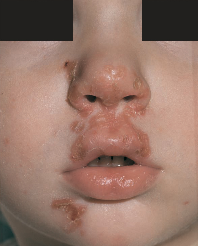
FIGURE 25-8 • Impetigo: MSSA Crusted erythematous erosions becoming confluent on the nose, cheek, lips, and chin in a child with nasal carriage of S. aureus.
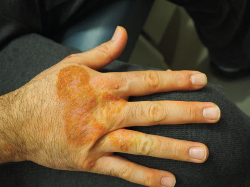
FIGURE 25-9 • Impetigo: MSSA Crusted, golden yellow confluent plaques on dorsal hand of adult with atopic dermatitis. Verrucae are present in the background.
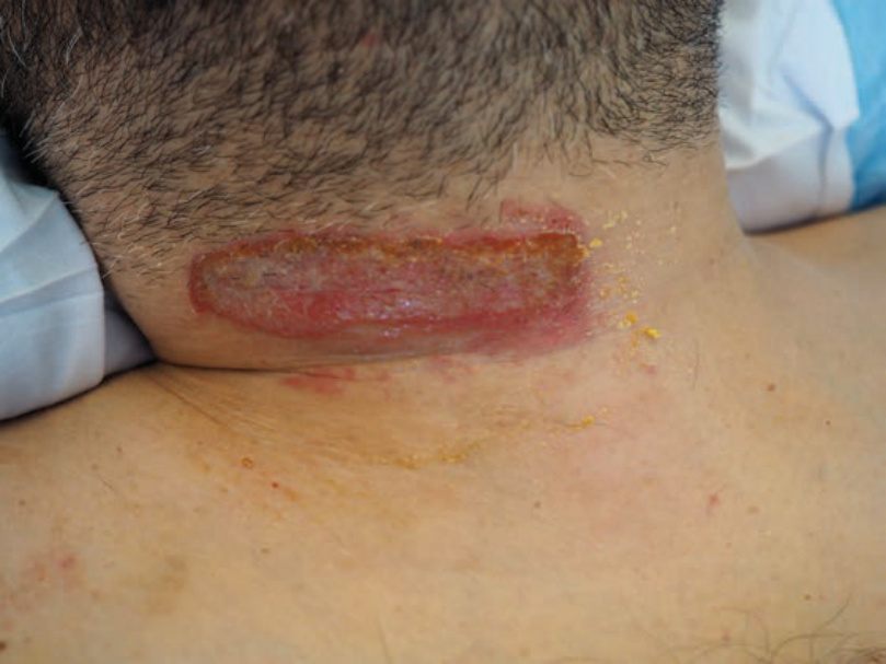
FIGURE 25-10 • Impetiginization of contact dermatitis: MRSA A 51-year-old man with golden yellow scale and crusts overlying a well demarcated moist red plaque after Neosporin use to a post-surgical wound.
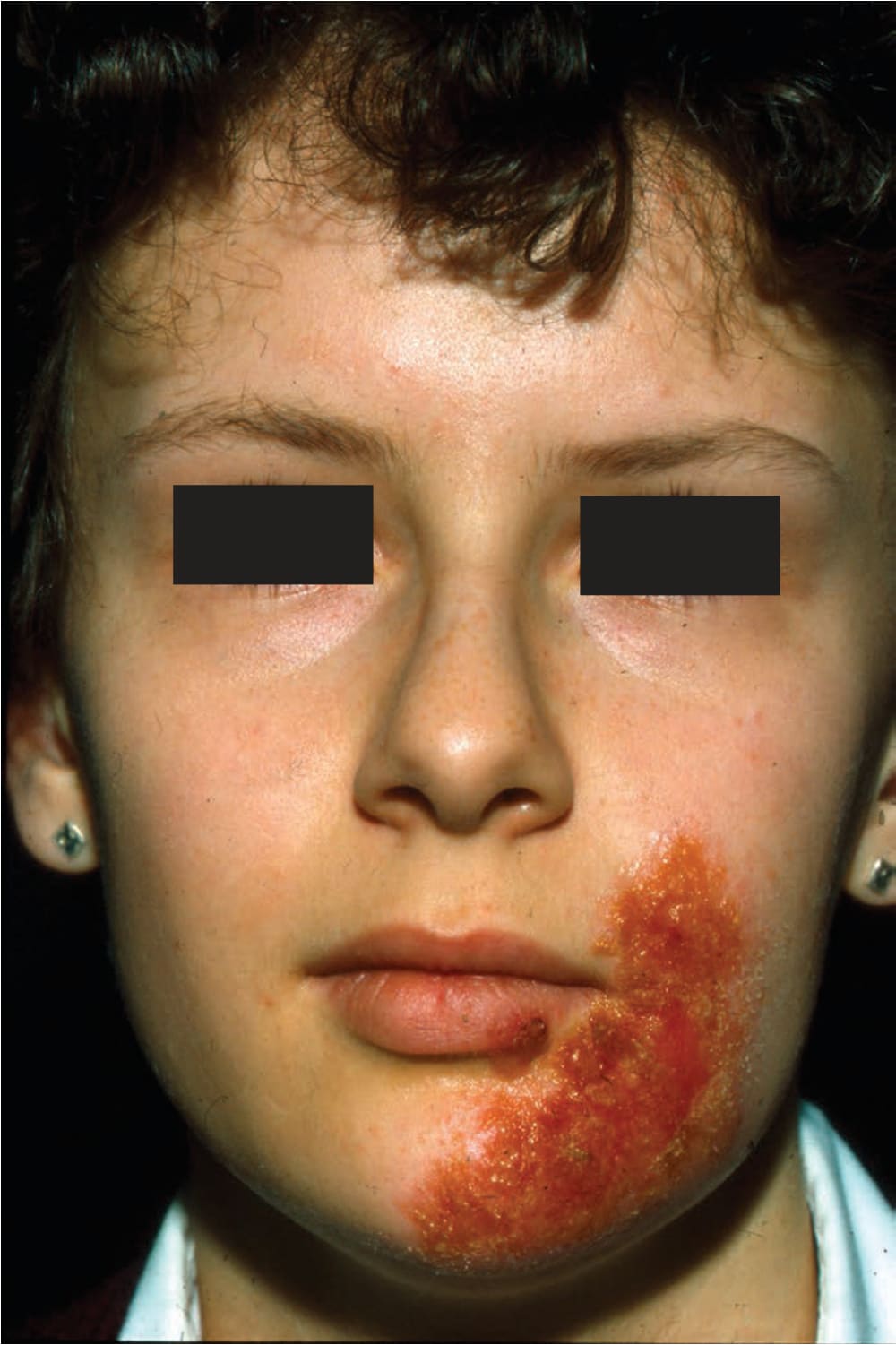
FIGURE 25-11 • Secondary infection of mild atopic dermatitis: MRSA An 11-year-old boy has yellowish crusted lesions on left cheek and chin.
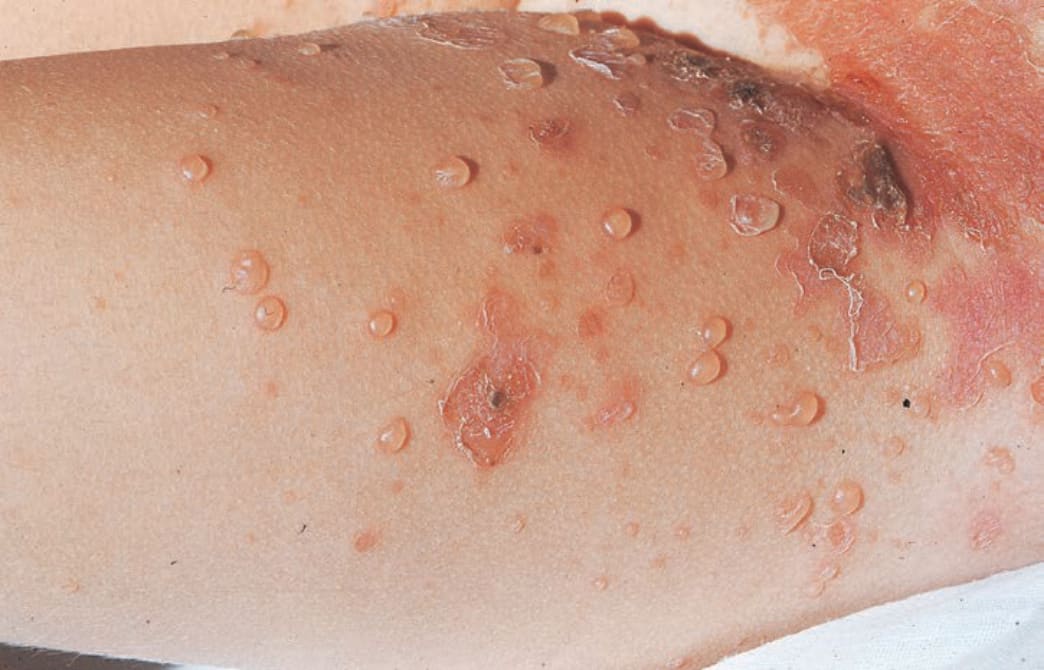
FIGURE 25-12 • Bullous impetigo Scattered, discrete, intact, and ruptured thin-walled blisters on the inguinal area and adjacent thigh of a child; lesions in the groin have ruptured, resulting in superficial erosions.
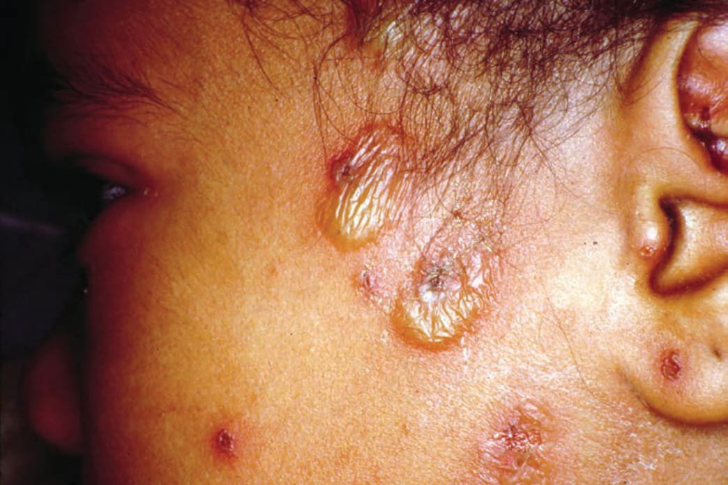
FIGURE 25-13 • Bullous impetigo: S. aureus Multiple flaccid bullae with yellowish exudate and surrounding punctate erythematous erosions in a child.
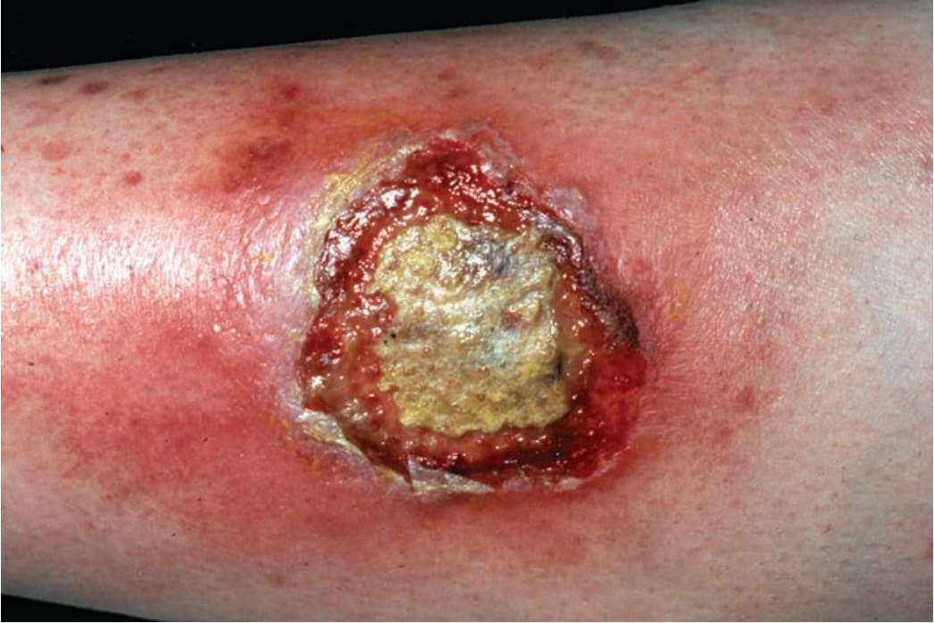
FIGURE 25-14 • Ecthyma: MSSA Thickly crusted ulcer on the leg that had been present on the lower leg of a homeless who had not taken off his boots for weeks. The crust was adherent and the site bled with debridement.