WOUND INFECTION
WOUND INFECTION
• Wound. Injury in which skin is surgically incised or traumatically injured (open wound) or in which blunt force trauma causes a contusion (closed wound). Wound infection: Skin and all wounds are colonized by bacteria and other microbes, i.e., cutaneous microbiome. Infection is characterized by pain, tenderness, purulence, erythema, warmth, and must be diagnosed on clinical as well as culture findings.
ETIOLOGY AND EPIDEMIOLOGY
CLASSIFICATION Traumatic wounds: Open or closed wounds (Fig. 25-36). Surgical wounds: Infection in surgical incisions (Fig. 25-37). Burn wounds: Burn wound may become superficially colonized with S. aureus; open burn-related surgical wound infection; burn wound cellulitis; invasive infection in debrided burn wounds (Fig. 25-38). Chronic ulcers: Arterial insufficiency; venous insufficiency;
neuropathic ulcers/diabetes mellitus; pressure ulcers (bedsores) (Figs. 25-39 to 25-41). Bites: Animal; human; insect. EPIDEMIOLOGY S. aureus in the most common pathogen in wound infections, MSSA, and increasingly MRSA. Surgical wound infection is up to 10 times more likely among patients who harbor S. aureus in nares. Hospital-acquired (nosocomial) or health-care–associated infections (most commonly surgical wound infections) are the most common complication affecting hospitalized patients. PATHOGENESIS Wounds are initially colonized by skin flora or introduced organisms. In some cases, these organisms proliferate, causing a host inflammatory response.
CLINICAL MANIFESTATION
LOCAL INFECTION Tenderness of wound area, erythema, hot, purulent drainage, and induration. Invasive infection: Malaise, anorexia, sweats, fever, or chills. Sepsis syndrome: Fever and hypotension. TYPES OF SURGICAL INFECTIONS Superficial infection of wound, wound infection with soft-tissue infection, i.e., cellulitis and erysipelas, soft-tissue abscess, necrotizing soft-tissue infection, and tetanus.
DIFFERENTIAL DIAGNOSIS
Allergic contact dermatitis (e.g., neomycin), pyoderma gangrenosum, and vasculitis.
DIAGNOSIS
Because all open wounds are colonized with microorganisms, diagnosis of infection relies on the clinical characteristics of the wound. Wound culture identifies the potential pathogen(s).
TREATMENT
Although all wounds require treatment, only infected lesions require antimicrobial therapy.
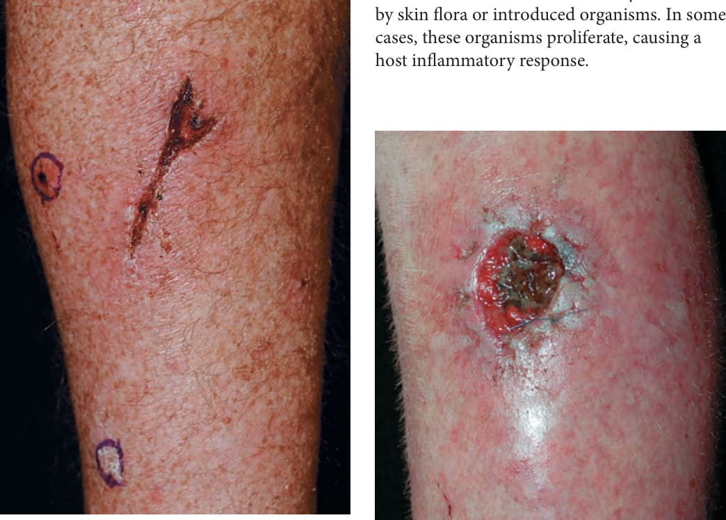
FIGURE 25-36 • Laceration infection in renal transplant recipient: MRSA A 60-year-old male immunosuppressed renal transplant recipient was unaware of a laceration on the calf. Erythema and induration are seen around the crusted wound. MRSA was isolated on culture. Two circled invasive squamous cell carcinomas are also seen on the calf.
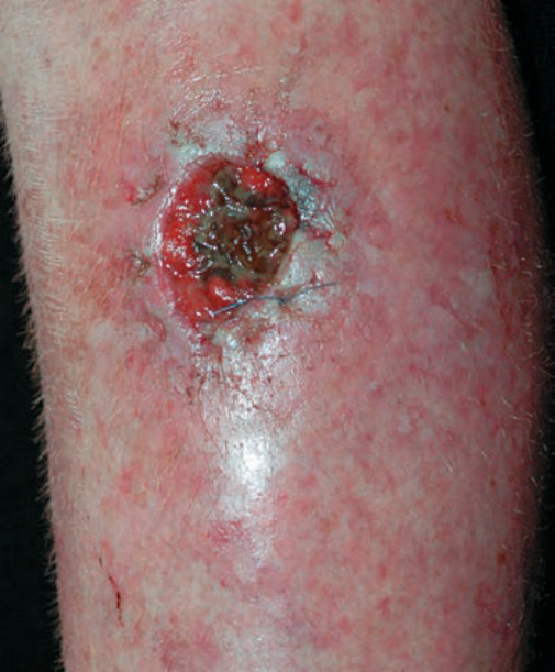
FIGURE 25-37 • Surgical excision wound infection: MSSA Surgical wound became painful and tender 7 days after excision of squamous cell carcinoma; soft tissue (cellulitis) is seen adjacent to the wound margin. Necrotic tissue is seen in the base.
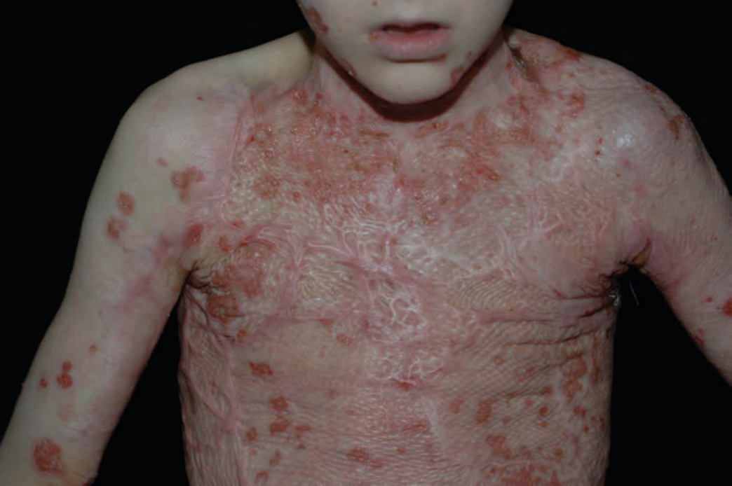
FIGURE 25-38 • Burn wound infection: MSSA A 10-year-old male with extensive third-degree thermal burn treated with autologous skin grafting has extensive new crusted erosions. MSSA was cultured from the infected site.
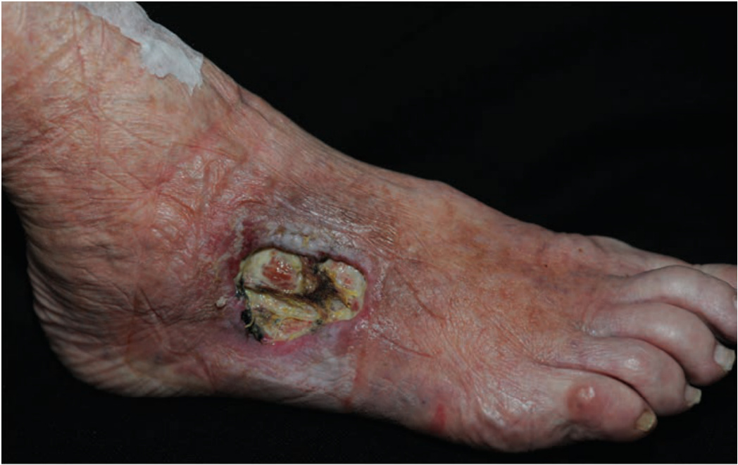
FIGURE 25-39 • Wound infection of stasis ulcer A 75-year-old woman with varicose veins and enlarging stasis ulcer infected with MRSA and Pseudomonas aeruginosa. IV antibiotics were administered. Incompetent veins were treated with endovascular laser ablation. The ulcer healed with minimal scar.
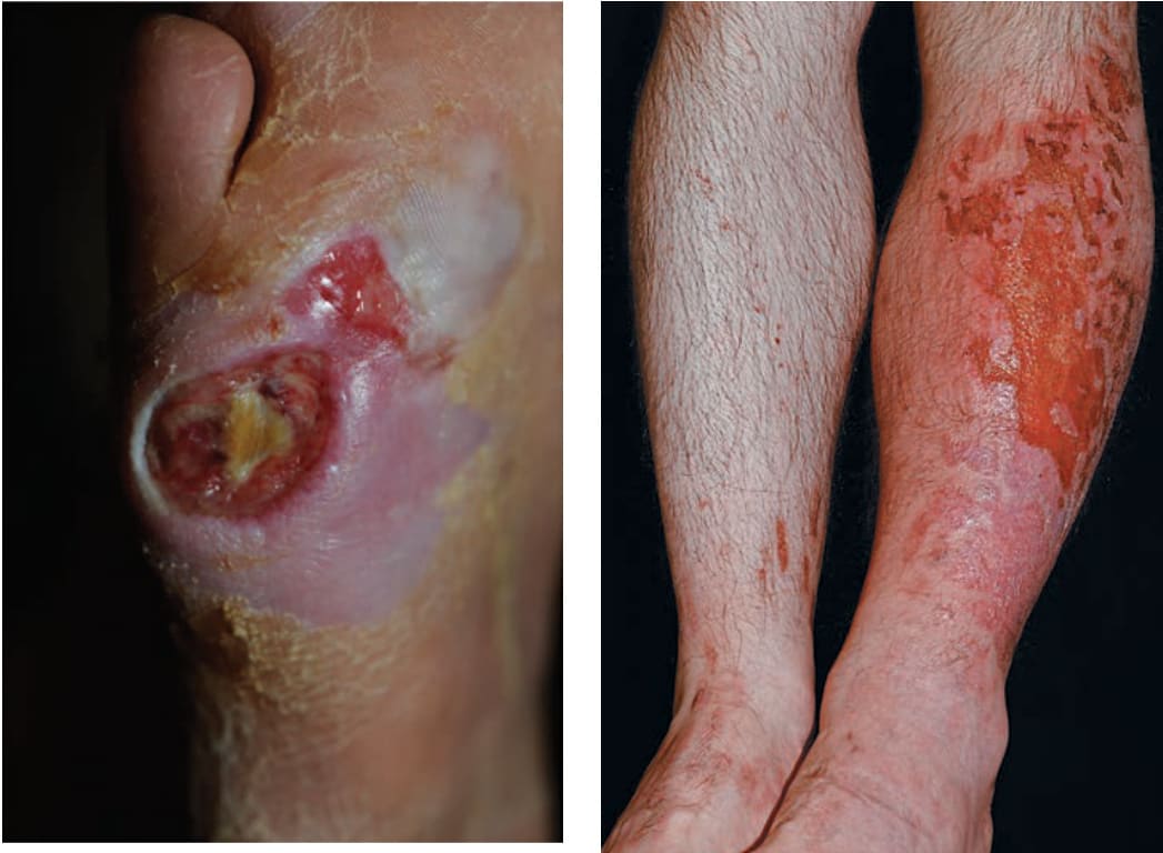
FIGURE 25-40 • Infection of diabetic ulcer: MRSA An 86-year-old man with diabetes mellitus type 2 had a chronic neuropathic ulcer on the R-lateral foot. The ulcer rapidly enlarged associated with fever and glucose of 450 mg/dL. MSSA was isolated from the wound. He was hospitalized and treated with IV antibiotics. He died 3 months later.