CUTANEOUS ANTHRAX
CUTANEOUS ANTHRAX ICD-10: A22.000
• Etiology. B. anthracis, a nonmotile, gram-positive, aerobic rod. Zoonosis. Spores can remain dormant in the soil for decades. Low-level germination occurs at the primary site, resulting in local edema and necrosis. Primary infection: Skin, pulmonary, and GI. Pathogenesis: Toxin mediated.
• Transmission. Zoonosis of mammals, especially herbivores. Human infections result from contact with contaminated wild and domestic animals or animal products. Human-to-human transmission does not occur. At risk: Farmers, herders; slaughterhouse, textile workers.
• Cutaneous anthrax. Accounts for 95% of anthrax cases in the United States.
• Cut or abrasion on exposed sites of head, neck, extremities. Nondescript, painless, pruritic papule (resembling insect bite) appears 3 to 5 days after introduction of endospores. In 1 to 2 days, evolves to vesicle(s) ± hemorrhage + necrosis. Vesicles rupture to form ulcers with extensive local edema (Fig. 25-46), ultimately forming dry eschars (1 to 3 cm). Satellite lesions can form in a nodular lymphangitis proximally on edematous extremity (Fig. 25-46).
• Differential Diagnosis, Ecthyma, brown recluse spider bite, ulceroglandular tularemia, orf, or glanders.
• Diagnosis. Isolation of B. anthracis from skin lesions, blood, or respiratory secretions, or by measuring specific antibodies in blood of persons with suspected symptoms.
• Course and Treatment. Mortality rate in untreated persons with cutaneous anthrax is about 20%. Systemic penicillin is the drug of choice; alternatives are erythromycin, azathioprin, clarithromycin, or cephalosporins.
A
B
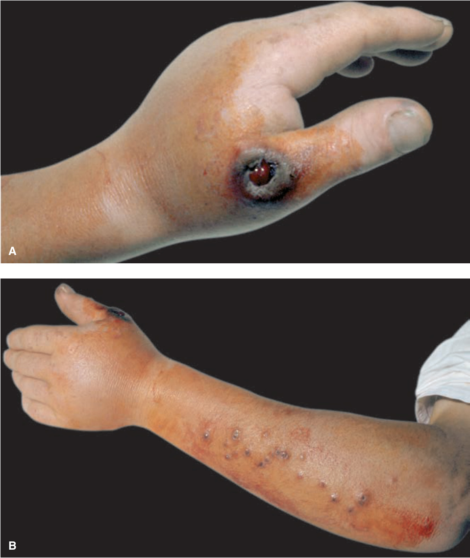
FIGURE 25-46 • A cutaneous anthrax A 40-year-old farmer with anthrax. (A) A black eschar at the site of inoculation with a central hemorrhagic ulceration on the thumb associated with massive edema of the hand. (B) A nodular lymphangitis extending proximally from the primary lesion on the thumb.