INFECTIVE ENDOCARDITIS
INFECTIVE ENDOCARDITIS ICD-10: I33
• Inflammation of endocardium. Infective and noninfective. Usually of heart valve. Characterized by vegetation that are made up of fibrin, platelets, and inflammatory cells (also microcolonies of microorganism if infective endocarditis).
• Infective endocarditis. Occurs at sites on altered endothelium or endocardium. The primary event is bacterial adherence to damaged valves during transient bacteremia. Bacteria grow within the cardiac lesion(s), i.e., vegetation, with local extension and cardiac damage. Subsequently, septic embolization occurs to skin, kidney, spleen, brain, etc. Circulating immune complexes may result in glomerulonephritis, arthritis, or various mucocutaneous manifestations of vasculitis. Embolization of vegetative fragments results in infection/infarction of remote tissues.
• Acute bacterial endocarditis rapidly damages cardiac structures, hematogenously seeds extracardiac sites, and may progress to death in a few weeks.
• Subacute bacterial endocarditis (SBE) causes structural damage slowly, rarely causes metastatic infection, and is gradually progressive unless complicated by a major embolic event or ruptured mycotic aneurysm.
• Noninfective endocarditis: Occurs on previously undamaged valves. Hypercoagulable state. Marantic endocarditis. Libman–Sacks endocarditis.
• Diagnosis: Based on clinical features, echocardiogram, and blood cultures.
CLINICAL MANIFESTATION
SEPTIC ARTERIAL EMBOLI Common with acute S. aureus endocarditis. Hematogenously seeded focal infection (Fig. 25-54). Apparent in up to 50% of patients. OSLER NODES Painful, erythematous nodules most commonly found on the pads of the
fingers and toes of some patients with infective endocarditis. JANEWAY LESIONS Nontender, erythematous, and nodular lesions most commonly found on the palms and soles (Fig. 25-55) of some patients with infective endocarditis.
SPLINTER HEMORRHAGES A small linear longitudinal subungual hemorrhage, initially red then brown. Middle third of nail bed in SBE. PETECHIAL LESIONS Small, nonblanching, reddish-brown macules. Occur on extremities, upper chest, mucous membranes (conjunctivae [Fig. 25-56], palate). Occur in crops. Fade after a few days (20% to 40%). ROTH SPOTS White spot in the retina close to the optic disk, often surrounded by hemorrhages; also seen in pernicious anemia and leukemia. SEPTIC EMBOLISM Painful, hemorrhagic macules, papules, or nodules, usually acral location.
COURSE AND TREATMENT
Varies with underlying cardiac disease and baseline health of the patient, as well as with the complications that occur. Complications: Congestive heart failure, stroke, other systemic embolization, or septic pulmonary embolization. Aortic valve involvement has a higher risk of death or need for surgery. Antibiotics.
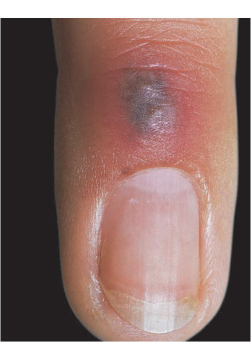
FIGURE 25-54 • Septic vasculitis associated with bacteremia Dermal nodule with hemorrhage and necrosis on the dorsum of a finger. This type of lesion occurs with bacteremia (e.g., S. aureus, gonococcus) and fungemia (e.g., Candida tropicalis).
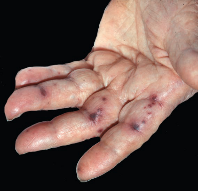
FIGURE 25-55 • Infective endocarditis, acute: Janeway lesions Hemorrhagic, infarcted papules on the volar fingers in a patient with S. aureus endocarditis.
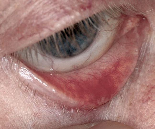
FIGURE 25-56 • Infective endocarditis, acute: Subconjunctival hemorrhage Submucosal hemorrhage of the lower eyelid in an elderly diabetic with enterococcal endocarditis; splinter hemorrhages in the midportion of the nail bed and Janeway lesions were also present on the volar fingers. Infection followed urosepsis.