SEPSIS
SEPSIS ICD: A40
• Sepsis is a whole-body inflammatory state, in response to infection. Can be complicated by multiple organ dysfunction.
• Characterized by fever or hypothermia, tachypnea, tachycardia, and, in severe cases, multiple organ dysfunction syndrome.
• Epidemiology. >1 million cases in the United States annually; >200,000 deaths. Two-thirds of cases occur in persons hospitalized for other illnesses. Incidence is increasing. Risk factors: Chronic disease and immunosuppression.
CLINICAL MANIFESTATION
Cutaneous infections as source of sepsis: Superficial skin infections, soft-tissue infections, wounds. E. gangrenosum (Fig. 25-32): P. aeruginosa most commonly. EXANTHEM See meningococcemia and RMSF (Fig. 25-50). PETECHIAE Cutaneous/oropharyngeal location suggests meningococcal infection; less commonly, H. influenzae. In patient with tick bite living in endemic area, RMSF (Figs. 25-51 and 25-52).
HEMORRHAGIC BULLOUS LESIONS V. vulnificus in patient (diabetes mellitus, liver disease) with history of eating raw oysters or clams (Fig. 25-33). Disseminated intravascular coagulation. See Section 20. Severe prolonged hypotension with acral necrosis of the fingers, hands, and feet (Fig. 25-57).
COURSE AND TREATMENT
Early sepsis is reversible; septic shock has high mortality/morbidity. High-dose antibiotics plus treatment of disseminated intravascular coagulation.
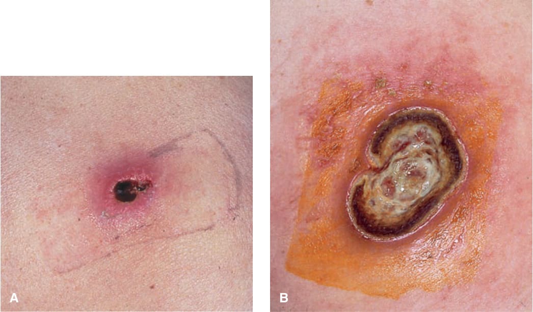
FIGURE 25-32 • Ecthyma gangrenosum of buttock: P. aeruginosa A 30-year-old male with HIV disease and neutropenia. (A) An extremely painful, infarcted area with surrounding erythema present for 5 days. This primary cutaneous infection was associated with bacteremia. (B) Two weeks later, the lesion had progressed to a large ulceration. The patient died 3 months later of P. aeruginosa pneumonitis associated with chronic neutropenia.
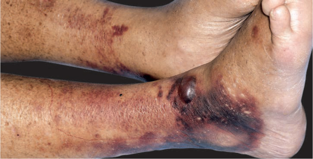
FIGURE 25-33 • Bilateral cellulitis of legs: V. vulnificus Bilateral hemorrhagic plaques and bullae on the legs, ankles, and feet of an older diabetic with cirrhosis. Unlike other types of cellulitis in which microorganisms enter the skin locally, this type of cellulitis (caused by V. vulnificus) usually follows a primary enteritis with bacteremia and subsequent dissemination to the skin. However, most cases initially diagnosed as bilateral cellulitis are inflammatory (eczema, stasis dermatitis, psoriasis) rather than infectious.
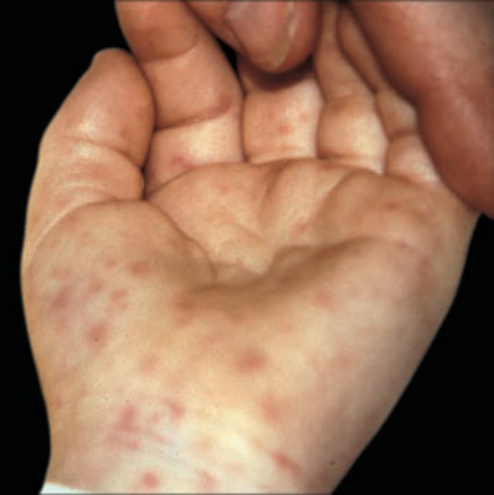
FIGURE 25-50 • Rocky Mountain spotted fever: Early Erythematous macules and papules appeared initially on the wrists of a young child. The lesions are not completely blanchable with pressure, indicating early hemorrhage of dermal blood vessels.
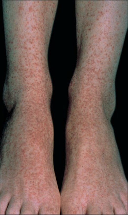
FIGURE 25-51 • Rocky Mountain spotted fever: Early Erythematous and hemorrhagic macules and papules appeared initially on the ankles of an adolescent.
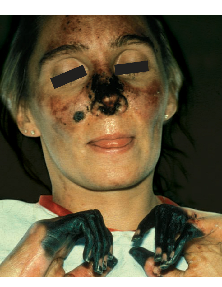
FIGURE 25-57 • Septic shock: Ischemic necrosis of acral sites Capnocytophaga canimorsus sepsis (dog bite) with prolonged hypotension and hypoperfusion resulted in infarction of fingers and nose.