MENINGOCOCCAL INFECTION
MENINGOCOCCAL INFECTION ICD-10: A39
• Etiology. N. meningitidis, colonizes nasopharynx. Infects only humans; no animal reservoirs. Spread by persons-to-person contact through respiratory droplets.
• Demography. The disease occurs sporadically throughout the world. The highest burden of the disease is caused by the cyclic epidemics occurring in the African “meningitis belt.”
CLINICAL MANIFESTATIONS
Small pink blanchable macules and papules occur soon after onset of disease (Fig. 25-58). With vascular friability and hemorrhage, petechiae and ecchymoses occur; first seen on the ankles, wrists, axillae, mucosal surfaces, and conjunctivae. A cluster of petechiae may be seen at pressure points, e.g., where a blood pressure cuff has been inflated. Ecchymoses and purpura may progress to hemorrhagic bullae, undergo necrosis, and ulcerate. Confluent necrotic hemorrhagic lesions may have bizarre-shaped, grayish to black necrosis (i.e., purpura fulminans) associated with
disseminated intravascular coagulation (DIC) in fulminant disease (Fig. 25-59). MENINGOCOCCEMIA SEPTICEMIA Meningococci enter the bloodstream and multiply, damaging the walls of the blood vessels and causing bleeding into the skin and organs. Characterized by development of shock and multiorgan failure. Peripheral gangrene may occur, requiring amputation in those who survive. WATERHOUSE–FRIDERICHSEN SYNDROME Fulminant meningococcal septicemia characterized by high fever, shock, widespread purpura, disseminated intravascular coagulation, thrombocytopenia, and adrenal insufficiency.
MENINGOCOCCAL MENINGITIS Bacteremia can result in the seeding of many organs, especially the meninges. The symptoms of meningococcal meningitis are those of typical bacterial meningitis, namely, fever, headache, stiff neck, and polymorphonuclear neutrophils (PMNs) in spinal fluid. CHRONIC MENINGOCOCCEMIA Intermittent bacteremia. Slow replication seeds various organs: Meninges, pericardium, large joints, and skin. Host inflammatory reaction limited to seeded site.
DIFFERENTIAL DIAGNOSIS
Adverse cutaneous drug eruptions, vasculitis, RMSF, and infective endocarditis.
DIAGNOSIS
Definitive etiologic diagnosis requires isolation of meningococci from blood or local site of infection.
COURSE
Onset of symptoms is sudden and death can follow within hours. In as many as 10% to 15% of survivors, there are persistent neurological defects, including hearing loss, speech disorders, loss of limbs, mental retardation, and paralysis.
TREATMENT
High-dose antibiotic therapy and treatment of DIC. PROPHYLAXIS Several vaccines are available to control the disease.
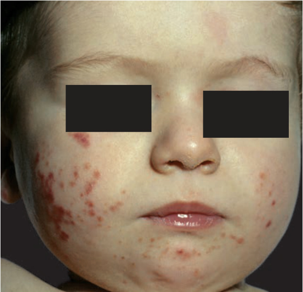
FIGURE 25-58 • Acute meningococcemia: Early exanthem Discrete, pink-to-purple macules and papules as well as purpura on the face of this young child. These lesions represent early disseminated intravascular coagulation with its cutaneous manifestation, purpura fulminans.
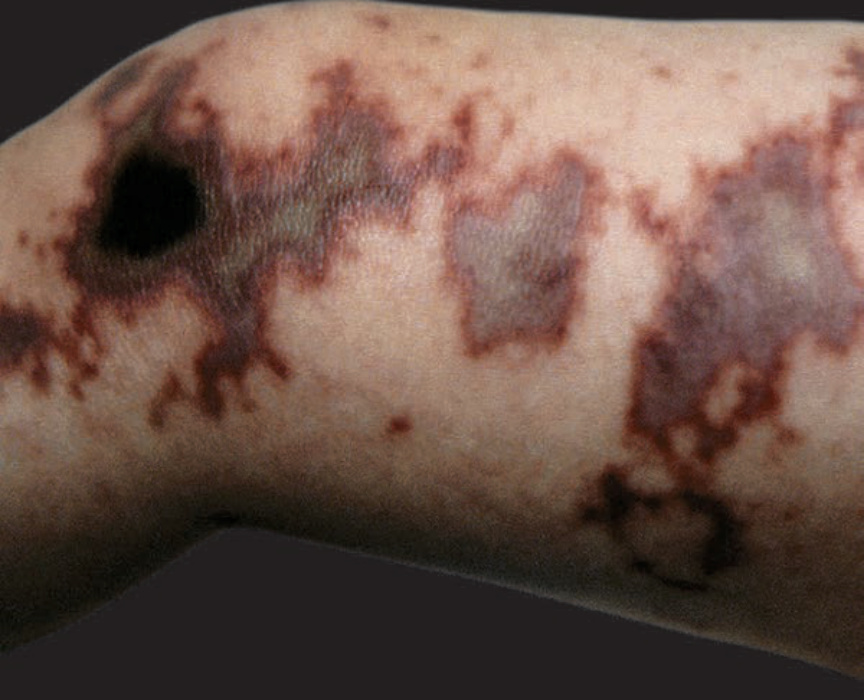
FIGURE 25-59 • Acute meningococcemia: Purpura fulminans Map-like, gray-to-black areas of cutaneous infarction of the leg in a child with NM meningitis and disseminated intravascular coagulation with purpura fulminans.