CUTANEOUS TUBERCULOSIS
CUTANEOUS TUBERCULOSIS ICD-10: A18.4
• Etiology. Mycobacterium tuberculosis complex. Commonly infects lungs; rarely skin.
• Transmission. Airborne spread of droplet nuclei from those with infectious pulmonary TB to lungs.
• Cutaneous Infection. Exogenous inoculation into skin. Direct extension from deeper tissues such as joint; lymphatic spread to skin; hematogenous spread to skin.
CLASSIFICATION
EXOGENOUS INOCULATION TO SKIN Primary inoculation tuberculosis (PIT), i.e., tuberculous chancre: Occurs at inoculated site in nonimmune host. Tuberculosis verrucosa cutis (TVC): Occurs at inoculated site in individual with prior tuberculosis infection. Tuberculosis may also result from bacille Calmette–Guérin (BCG) immunization. ENDOGENOUS SPREAD TO SKIN Lymphatics, hematogenous, and bodily fluids (sputum, feces, or urine). Lupus vulgaris. Scrofuloderma. Metastatic tuberculosis abscess. Acute miliary tuberculosis. Orificial tuberculosis.
PATHOGENESIS
Type of clinical lesion depends on route of cutaneous inoculation and immunologic status of the host.
• Cutaneous inoculation results in a tuberculous chancre in the nonimmune host and TVC in the immune host.
• Direct extension from underlying tuberculous infection, i.e., lymphadenitis or tuberculosis of bones and joints, results in scrofuloderma.
• Lymphatic spread to skin results in lupus vulgaris.
• Hematogenous dissemination results in acute miliary tuberculosis, lupus vulgaris, or metastatic tuberculosis abscess.
• Autoinoculation from body fluids such as sputum, urine, and feces results in orificial tuberculosis.
Globally, the incidence of cutaneous tuberculosis is increasing, associated with HIV disease. Problem of multidrug resistance (MDR) is also common in persons with HIV disease.
CLINICAL MANIFESTATION
PIT Initially, papule occurs at the inoculation site 2 to 4 weeks after inoculation. Lesion enlarges to a painless ulcer, tuberculous chancre (Fig. 25-68) with shallow granular base.
Older ulcers become indurated with thick crusts. Deeper inoculation results in subcutaneous abscess. Most common on exposed skin at sites of minor injuries. Oral ulcers on gingiva or palate occur after ingestion of bovine bacilli in nonpasteurized milk. Regional lymphadenopathy occurs several weeks after appearance of the ulcer (chancriform syndrome) (Fig. 25-68). TVC Initial papule with violaceous halo. Evolves to hyperkeratotic, warty, or firm plaque (Fig. 25-69). Clefts and fissures occur from which pus and keratinous material can be expressed. Border often irregular. Lesions are usually single, but multiple lesions occur. Most commonly on the dorsolateral hands and fingers. In children, on the lower extremities, knees. No lymphadenopathy. LUPUS VULGARIS Initial papule ill-defined and soft, evolves into well-defined, irregular plaque (Fig. 25-70). Reddish-brown. Diascopy (glass slide pressed against skin) shows an “apple jelly” color (i.e., orange-tan). Lesions
are characteristically soft and friable. Surface is initially smooth or slightly scaly but may become hyperkeratotic. Hypertrophic forms result in soft tumorous nodules. Ulcerative forms present as punched-out, often serpiginous ulcers surrounded by soft, brownish infiltrate. Usually solitary, but several sites may occur. Most lesions on the head and neck, most often on nose, ears, or scalp. Lesions on ears or nose can result in destruction of underlying cartilage. Scarring is prominent. Characteristically new brownish infiltrates occur within atrophic scars. SCROFULODERMA Firm subcutaneous nodule that initially is freely movable; lesion then becomes doughy and evolves into irregular, deep-seated node or plaque that liquefies and perforates (Fig. 25-71). Ulcers and irregular sinuses, usually of linear or serpiginous shape, discharge pus or caseous material. Edges are undermined, inverted, with dissecting subcutaneous pockets alternating with soft, fluctuating infiltrates and bridging scars. Most often occurs
in the parotid, submandibular, and supraclavicular regions; lateral neck; scrofuloderma most often results from contiguous spread from affected lymph nodes or tuberculous bones (phalanges, sternum, or ribs) or joints. METASTATIC TUBERCULOSIS ABSCESS Subcutaneous abscess, nontender, “cold,” and fluctuant. Coalescing with overlying skin, breaking down and forming fistulas and ulcers. Single or multiple lesions, often at sites of previous trauma. ACUTE MILIARY TUBERCULOSIS Exanthem. Disseminated lesions are minute macules and papules or purpuric lesions. Sometimes vesicular and crusted. Removal of crust reveals umbilication. Disseminated on all parts of the body, particularly the trunk. ORIFICIAL TUBERCULOSIS Small yellowish nodule on mucosa breaks down to form painful circular or irregular ulcer (Fig. 25-72) with undermined borders. Surrounding mucosa swollen, edematous, and inflamed. Since orifical tuberculosis results from autoinoculation of mycobacteria from progressive tuberculosis of internal organs, it is usually found on the oral, pharyngeal (pulmonary tuberculosis), vulvar (genitourinary tuberculosis), and anal (intestinal tuberculosis) mucous membranes. Lesions may be single or multiple, and in the
mouth most often occur on the tongue, soft and hard palate, or lips.
DIAGNOSIS
Clinical findings, tuberculin skin testing (Fig. 25-73), dermatopathology, confirmed by
isolation of M. tuberculosis on culture or by PCR.
COURSE
The course of cutaneous tuberculosis is quite variable, depending on the type of cutaneous infection, amount of inoculum, extent of extracutaneous infection, age of the patient, immune status, and therapy.
TREATMENT
Only PIT and TVC are limited to the skin. All other patterns of cutaneous tuberculosis are associated with systemic infection that has disseminated secondarily to skin. As such, therapy should be aimed at achieving a cure, avoiding relapse, and preventing emergence of drug-resistant mutants. ANTITUBERCULOUS THERAPY Prolonged antituberculous therapy with at least two drugs is indicated for all cases of CTb except for TVC that can be excised.
• Standard antituberculous therapy:
• Isoniazid (5 mg/kg daily).
• Rifampin (600 mg/kg daily).
• Supplemented in initial phases with:
• Ethambutol (25 mg/kg daily) and/or
• Streptomycin (10 to 15 mg/kg daily) and/or
• Pyrazinamide (15 to 30 mg/kg daily).
Isoniazid and rifampin for at least 9 months; can be shortened to 6 months if four drugs are given during the first 2 months. MULTIDRUG RESISTANT (MDR) TB Incidence is increasing.
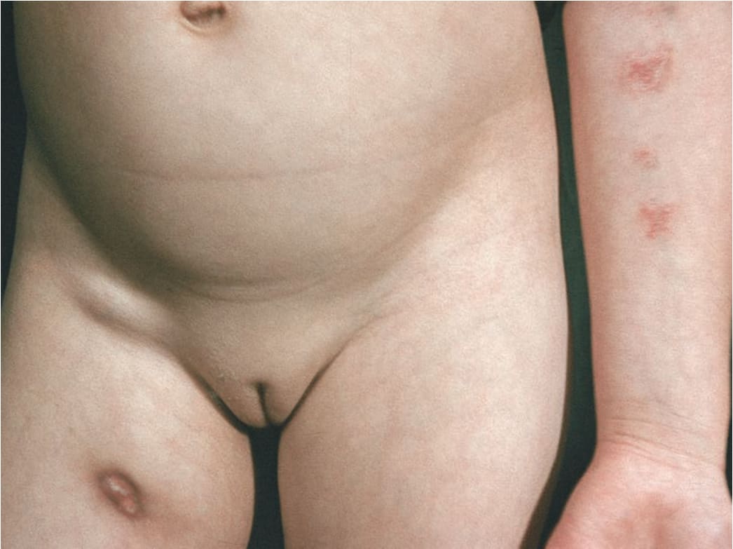
FIGURE 25-68 • Primary inoculation tuberculosis A large, ulcerated nodule at the site of Mycobacterium tuberculosis inoculation on the right thigh associated with inguinal lymphadenopathy. The erythematous papules on the left forearm occurred at the site of tuberculin testing.
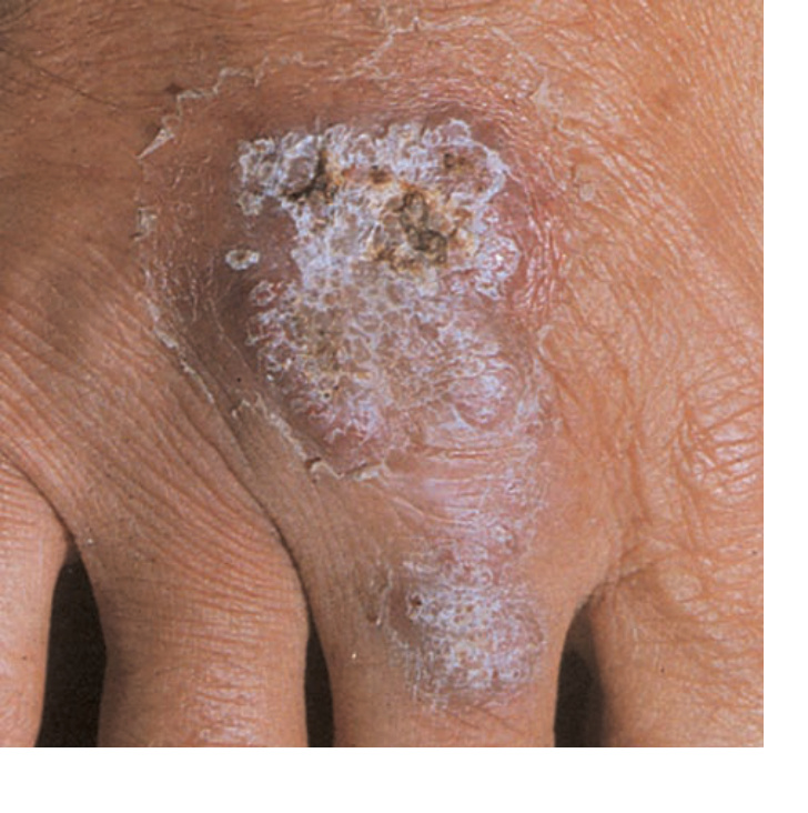
FIGURE 25-69 • Tuberculosis verrucosa cutis A 40-year-old male with warty and crusted plaques on the dorsum of the hand for 6 months. (Reproduced with permission from Sethi A. Tuberculosis and infections with atypical Mycobacteria. In: Goldsmith LA, Katz SI, Gilchrest BA, et al., eds. Fitzpatrick’s Dermatology in General Medicine. 8th ed. New York, NY: McGraw Hill; 2012.)
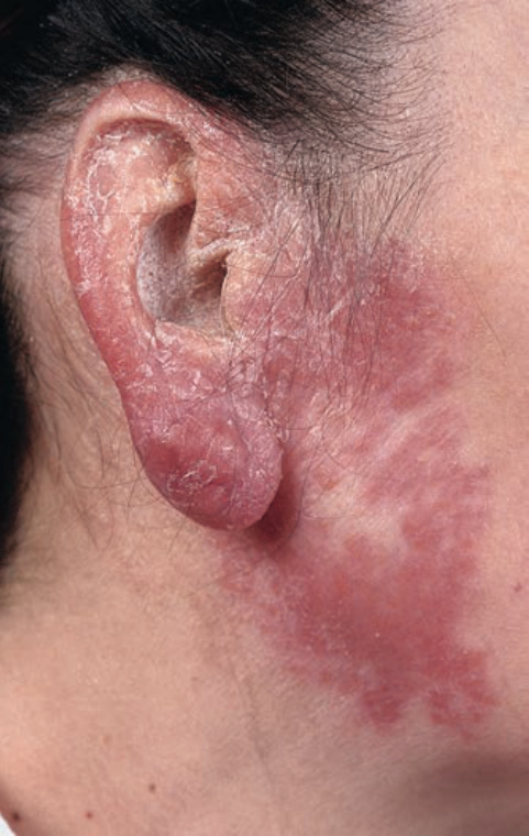
FIGURE 25-70 • Lupus vulgaris Reddish-brown plaque, which on diascopy exhibits the diagnostic yellow-brown apple-jelly color. Note nodular infiltration of the earlobe, scaling of the helix, and atrophic scarring in the center of the plaque.
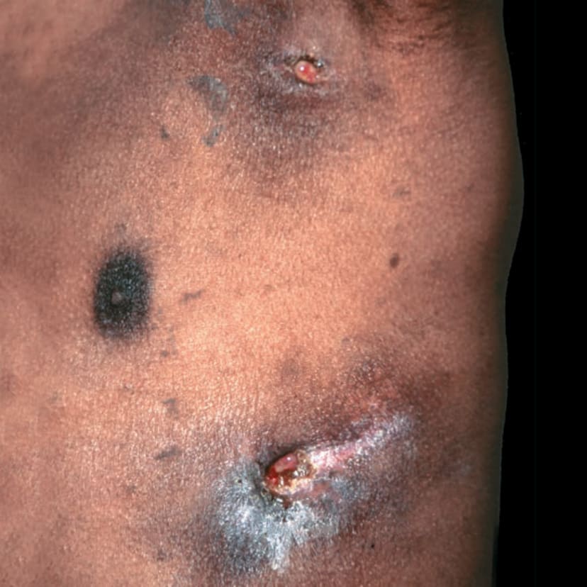
FIGURE 25-71 • Scrofuloderma: Lateral chest wall Two ulcers on the chest wall and axilla are associated with underlying sinus tracts.
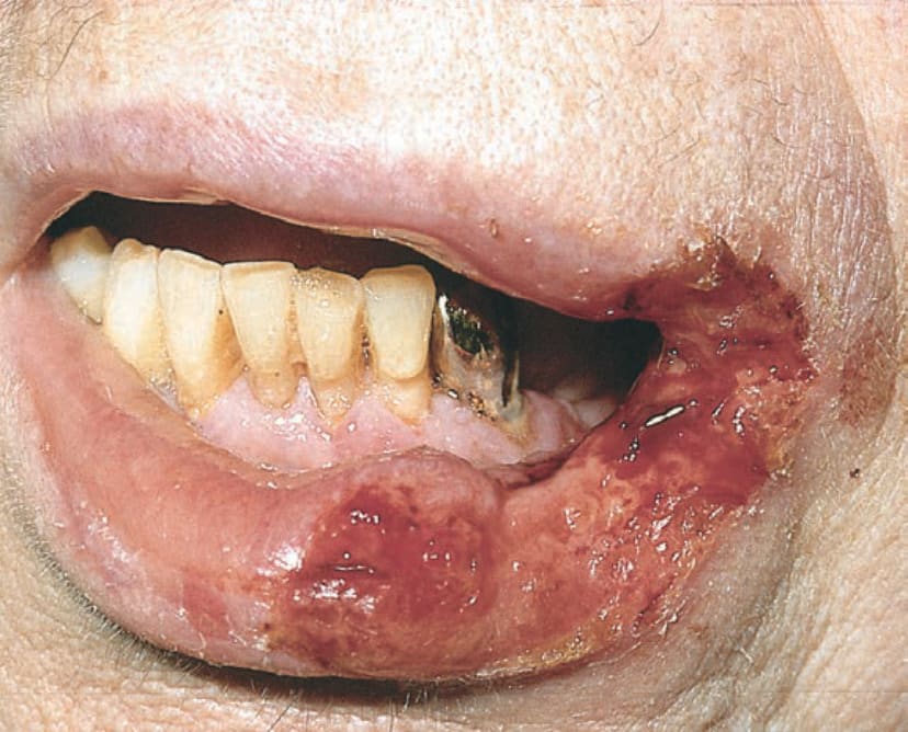
FIGURE 25-72 • Orificial tuberculosis: Lips A large, very painful ulcer on the lips of this patient with advanced cavitary pulmonary tuberculosis.
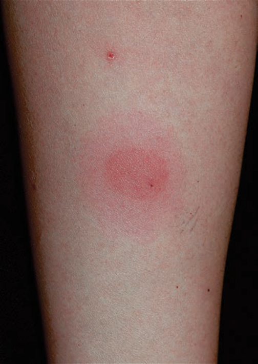
FIGURE 25-73 • Purified protein derivative or Mantoux test: Positive test A 31-year-old Taiwanese female with psoriasis, with a negative skin test 1 year previously, was retested prior to beginning etanercept. She had become infected while visiting her father, who had pulmonary tuberculosis, in Taiwan. A red plaque with surrounding erythema is seen at the test site.