CUTANEOUS AMEBIASIS
CUTANEOUS AMEBIASIS ICD-10: A06.7
Amebiasis is caused by Entamoeba histolytica, which infects the GI tract and rarely the skin.
• Incidence. ~50 million cases globally infected with Entamoeba. Majority of infections caused by noninvasive E. dispar. 10% of those colonized with E. histolytica develop amebic colitis. More prevalent in tropics and in rural areas; inadequate sanitation and crowding. Skin involvement is associated with malnutrition and immunocompromise (HIV/AIDS and solid organ transplantation).
CLINICAL MANIFESTATIONS
Cutaneous amebiasis begins as an indurated pustule that evolves to a painful ragged ulcer, foul smelling, and covered with pus or necrotic debris (Fig. 29-8). Usually, a consequence of underlying amebic abscess invading skin. Typical sites are perianal area (extension of sigmorectal involvement) (Fig. 29-8) or abdominal wall (draining sinus from liver or colon). Penis or vulva may become infected during intercourse. Surgical wound infections may follow removal of hepatic or abdominal abscess. Remote ulcers (e.g., face) may result from autoinoculation.
COURSE AND TREATMENT
Without treatment lesion progressively enlarges. Treat with sulfadiazine and pyrimethamine, clindamycin.
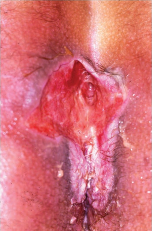
FIGURE 29-8 • Perianal ulcer in a 35-year-old man, 4 weeks after removal of condylomata acuminata by electrodesiccation The ulcer showed no tendency to heal and a biopsy of its base revealed an inflammatory infiltrate with oval protozoa with phagocytosed red blood cells. Real-time PCR was positive for entameba histolytica. (Reproduced with permission from Posch C, Walochnik J, Schuller-Lukic B, et al. Perianal ulcer-amebiasis cutis. J Dtsch Dermatol Ges. 2011;9(8):649–650. © The authors. Journal compilation © Blackwell Verlag GmbH, Berlin.)