GENITAL WARTS
GENITAL WARTS
CLINICAL MANIFESTATION
Usually asymptomatic, except for cosmetic appearance. Anxiety of having STI. Obstruction if large mass.
A
B
MUCOCUTANEOUS LESIONS Four clinical types of genital warts occur: Small papular (Fig. 30-1). Condyloma acuminatum. Cauliflower-floret (acuminate or pointed) lesions (Figs. 30-2 to 30-4).
A B
Keratotic warts (Figs. 30-5 and 30-6). Flat-topped papules/plaques (most common on cervix) (Fig. 30-7). Skin-colored, pink, red, tan, and brown. Solitary, scattered, and isolated, or they form voluminous confluent masses. In immunocompromised individuals, lesions may be huge (Fig. 30-5). Sites of predilection. Male: Frenulum, corona, glans penis, prepuce, shaft (Figs. 30-1 and 30-3), and scrotum. Female: Labia, clitoris, periurethral area, perineum, vagina, and cervix (flat lesions) (Fig. 30-7). Both sexes: Perineal, perianal (Fig. 30-5), anal canal, rectal; urethral meatus, urethra, and bladder; oropharynx.
Laryngeal Papillomas
• Relatively uncommon; associated with HPV-6 and -11.
• Arise most commonly on true vocal cords of the larynx.
• Age: Children <5 years of age, adults >20 years of age.
• Risk of squamous cell carcinoma in situ (SCCIS) and invasive squamous cell carcinoma (SCC).
DIFFERENTIAL DIAGNOSIS
PAPULAR/NODULAR EXTERNAL GENITAL LESIONS Normal anatomy (e.g., sebaceous glands, pearly penile papules, and vestibular papillae), squamous intraepithelial lesions (SILs), SCCIS, invasive SCC, benign neoplasms (moles, seborrheic keratoses, skin tags, pilar cyst, and angiokeratoma), inflammatory dermatoses (lichen nitidus, lichen planus), molluscum contagiosum, condylomata lata, folliculitis, and scabetic nodules.
LABORATORY EXAMINATIONS
PAP SMEAR Encourage all women to have an annual Pap smear since HPV is the major etiologic agent for cancer of the cervix. Anal Pap test with a cervical brush and fixative solution helps detect anal dysplasia. DERMATOPATHOLOGY Biopsy is indicated if diagnosis is uncertain; lesions do not respond to standard therapy and worsen during therapy; the patient is immunocompromised; warts are pigmented, indurated, fixed, and/or ulcerated. Indicated in some cases to confirm
A
B C
diagnosis and/or rule out SCCIS or invasive SCC. DETECTION OF HPV DNA Presence of HPV DNA and specific HPV types determined on smears and lesional biopsy by in situ hybridization.
Serology Genital warts are markers of unsafe sexual practices, and patients should be screened for other STDs.
DIAGNOSIS
Clinical diagnosis, occasionally confirmed by biopsy.
COURSE
HPV is highly infectious, with an incubation period of 3 weeks to 8 months. Most HPV-infected individuals who develop genital warts do so 2 to 3 months after becoming infected. If left untreated, genital warts may resolve on their own, remain unchanged, or grow. After regression, subclinical infection may persist for life. Recurrence may occur with normal immune function as well as in immunocompromised. Recurrences more commonly are reactivation of subclinical infection than
reinfection. In pregnancy, genital warts may increase in size and number, show increased vaginal involvement, and have an increased rate of secondary bacterial infection. Children delivered vaginally of mothers with genital HPV infection are at risk for developing recurrent respiratory papillomatosis in later life. HPV types 16, 18, 31, and 33 are the major etiologic factors for in situ and invasive SCC: Cervix; external genitalia (vulva and penis); anus and perineum.
MANAGEMENT
PREVENTION Use of condoms reduce transmission. HPV vaccine protects against nine strains of HPV and cervical cancer later in life. GOAL OF TREATMENT Removal of exophytic warts and reduction of signs and symptoms. No therapy has been shown to eradicate HPV or prevent cervical or anogenital cancer. Treatment is more successful if warts are small and present for <1 year. Risk of transmission might be reduced by “debulking” genital warts.
A
B
SELECTION OF TREATMENT Guided by preference of patient—avoid expensive therapies, toxic therapies, and procedures that result in scarring. See Section 27. PATIENT APPLIED AGENTS Imiquimod 5% cream, podophylox 0.5% solution. CLINICIAN ADMINISTERED THERAPY Cryosurgery, podophyllin 10% to 25%, trichloroacetic acid 80% to 90%, surgical removal, and electrodesiccation. Intralesional Cidfovoir.
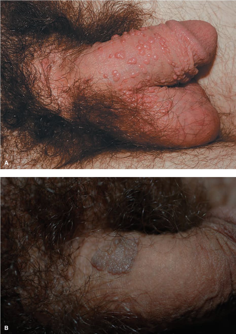
FIGURE 30-1 • Papular warts: penis (A) A 23-year-old man with penile lesions for 6 months. Multiple skin-colored papules on the penis and scrotum. (B) A 28-year-old man with skin-colored pebbled smooth papules.
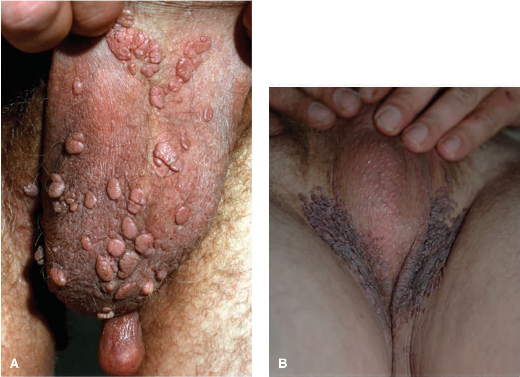
FIGURE 30-2 • Condyloma acuminata (A) A 35-year-old man with cluster of warts on the scrotum for 2 months. (B) A 22-year-old man with clusters of soft warts on the inguinal canals adjacent to the scrotum.
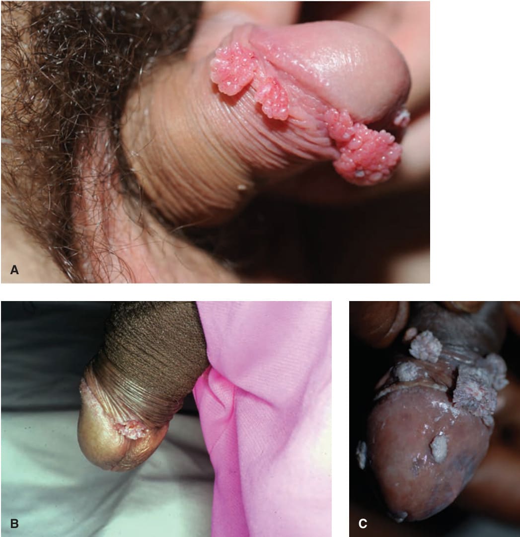
FIGURE 30-3 • Condylomata acuminata: penis (A) A 20-year-old male with Crohn disease treated with infliximab infusion. Condylomata on the distal foreskin resemble cauliflower floret-like papules. (B) Filiform papules on the ventral glans penis. (C) Multiple cauliflower floret-like papules in a 40-year-old man with HIV.
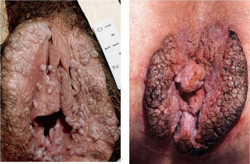
FIGURE 30-4 • Condylomata acuminata: vulva Multiple, pink-brown, soft papules on the labia.
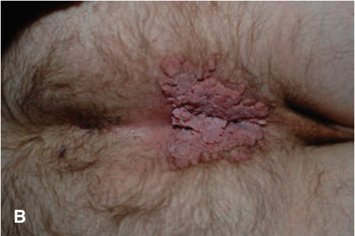
FIGURE 30-5 • (A,B) Genital warts Two different men, both with HIV disease and hyperkeratotic condylomata acuminata seen on the anal and perineal area.
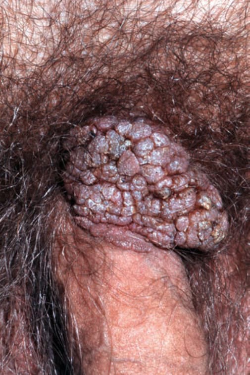
FIGURE 30-6 • Keratotic external genital warts (EGW): male A 51-year-old male with lesions at the base of penis for several years. Lesional biopsy reported EGW ruling out verrucous carcinoma.
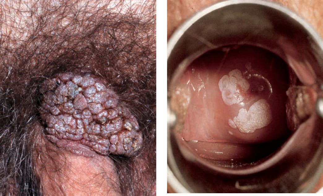
FIGURE 30-7 • Condylomata acuminata: uterine cervix Sharply demarcated, whitish, and flat plaques becoming confluent around the cervix.