LYMPHOGRANULOMA VENEREUM
LYMPHOGRANULOMA VENEREUM ICD-10: A55
• Clinical manifestations depend on the site of entry of C. trachomatis (the sex contact site) and the stage of disease progression: Inguinal syndrome, rectal syndrome, and pharyngeal syndrome.
ETIOLOGY AND EPIDEMIOLOGY
ETIOLOGY C. trachomatis, obligate intracellular bacterium. Major outer-membrane protein delineates >20 serovars (immunotypes): Trachoma: Serovars A, B, Ba, and C. Mucosal STDs: Serovars D-K (most common bacterial STD). Invasive STD: Serovars L1, L2, L3 (in United States, L2 most commonly). TRANSMISSION Sexual: C. trachomatis in purulent exudate is inoculated onto skin or mucosa of sexual partner and gains entry through minute lacerations and abrasions. Perinatal. Heterosexual men: Acute infection presents as
inguinal syndrome. Women/homosexual men: Anogenitorectal syndrome most common. PREVALENCE Chlamydial urethritis more common in heterosexual men and high socioeconomic status. Prevalence of cervical infection in the United States: 5% for asymptomatic college students; >10% in family planning clinics: >20% in STD clinics. PATHOGENESIS Primarily an infection of lymphatics and lymph nodes. Lymphangitis and lymphadenitis occur in the drainage field of the inoculation site with subsequent perilymphangitis and periadenitis. Necrosis occurs; loculated abscesses, fistulas, and sinus
tracts develop. As the infection subsides, fibrosis replaces acute inflammation with resulting obliteration of lymphatic drainage, chronic edema, and stricture.
CLINICAL MANIFESTATION
ACUTE LYMPHOGRANULOMA VENEREUM Primary genital lesion noticed in less than one-third of men and rarely in women. In heterosexual men and women: Small painless vesicle or nonindurated ulcer/papule on the penis or labia/posterior vagina/fourchette; heals in a few days. With receptive anal intercourse, primary anal or rectal infection develops after receptive anal intercourse. Infection can spread from the primary site of infection to regional lymphatics. Papule, shallow erosion or ulcer, grouped small erosions or ulcers (herpetiform), or nonspecific urethritis. Cordlike lymphangitis of dorsal penis may follow. Lymphangial nodule (bubonulus) may rupture, resulting in sinuses and fistulas of the urethra and deforming scars of the penis. Multilocular suppurative lymphadenopathy. Cervicitis, perimetritis, salpingitis may occur. Receptive anal intercourse: Primary anal rectal infection (hemorrhagic proctitis with regional lymphadenitis). Erythema nodosum in 10% of cases (see Section 7). INGUINAL SYNDROME Characterized by painful inguinal lymphadenopathy beginning 2 to 6 weeks after presumed exposure. Unilateral in two-thirds of cases; palpable iliac/femoral nodes often present on same side (Fig. 30-35). Initially, nodes are discrete, but progressive periadenitis results in a matted mass of nodes that may become fluctuant and suppurative. Overlying skin becomes fixed, inflamed, thin, and eventually develops multiple draining fistulas. Groove sign: Extensive enlargement of
chains of inguinal nodes above and below the inguinal ligament (Fig. 30-35). Unilateral bubo in two-thirds of cases (most common presentation) (Fig. 30-35). Marked edema and erythema of skin overlying node. One-third of inguinal buboes rupture; two-thirds slowly involute. Seventy-five percent of cases have deep iliac node involvement with a pelvic mass that seldom suppurates. Anogenitorectal syndrome associated with receptive anal intercourse, proctocolitis, hyperplasia of intestinal and perirectal lymphatic tissue. Resultant abscesses, fistulas, and rectal stricture. Overgrowth of lymphatic tissue results in lymphorrhoids (resembling hemorrhoids) or perianal condylomata. ESTHIOMENE Elephantiasis of genitalia, usually females, which may ulcerate, occurring 1 to 20 years after primary infection.
DIFFERENTIAL DIAGNOSIS
PRIMARY STAGE GH, primary syphilis, and chancroid. INGUINAL SYNDROME Incarcerated inguinal hernia, plague, tularemia, tuberculosis, GH, syphilis, chancroid, and lymphoma.
DIAGNOSIS
Diagnosis is based on clinical findings. Exclude other causes of inguinal lymphadenopathy or genital ulcers.
COURSE
Highly variable. Bacterial secondary infections may contribute to complications. Rectal stricture is late complication. Spontaneous remission is common.
TREATMENT
Oral doxycycline 100 mg twice daily for 21 days or oral erythromycin base 500 mg four times daily for 21 days.
A
B
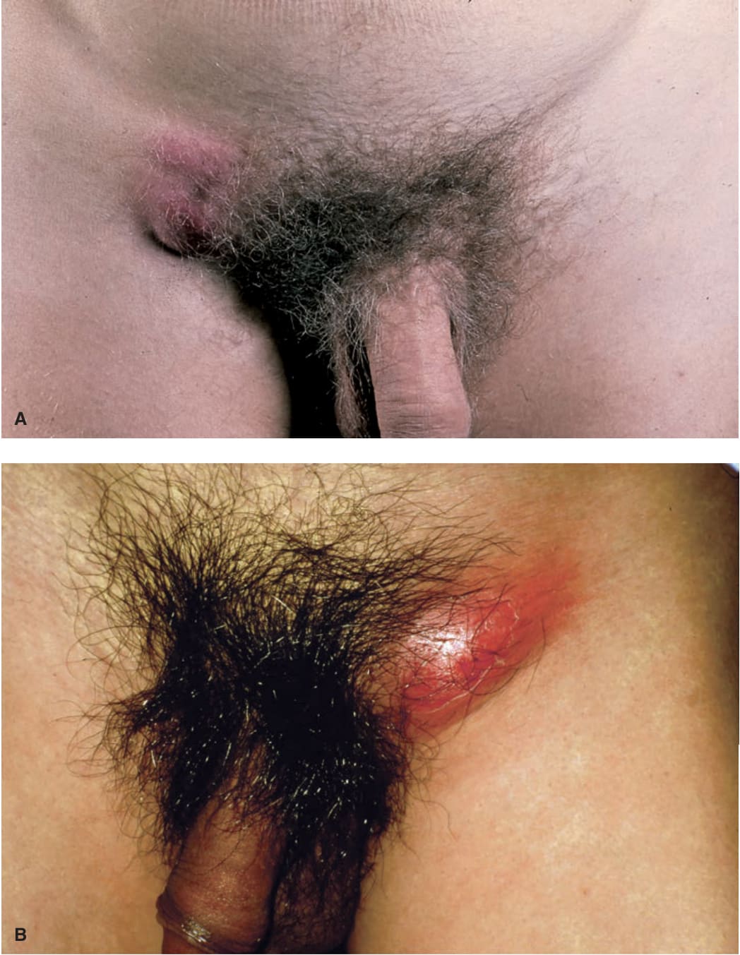
FIGURE 30-35 • Lymphogranuloma venereum: Groove sign Striking tender lymphadenopathy occurring at the right (A) and left (B) femoral and inguinal lymph nodes separated by a groove made by Poupart ligament (groove sign).