LEUKOPLAKIA
LEUKOPLAKIA ICD-10: K13.21
• Leukoplakia is a chronic white plaque/lesion in the oropharynx.
• Premalignant leukoplakia has histologic atypia.
• Leukoplakia is a descriptive clinical term regarding morphology: Squamous cell carcinoma, in situ and invasive, must be ruled out.
• Findings: A white plaque that cannot be wiped off and cannot be diagnosed as any other distinct lesion and may be premalignant or malignant.
• Definitive diagnosis should be made on clinical findings and histology.
• When diagnosis is definitive histologically, “leukoplakia” is no longer appropriate. The differential diagnosis includes SCCIS, invasive SCC, candidiasis, migratory glossitis, radiotherapy and chemotherapy-induced mucositis, lichen planus, and lupus erythematosus.
The differential diagnosis of leukoplakia is shown in Table 33-1.
Lesion/Disorder Characteristics
Leukoedema (Fig. 33-12) Grayish-white opalescence of buccal mucosa; variant of normal. Histology: Aacanthosis.
Frictional keratosis/ lichen simplex chronicus (Fig. 33-13)
Keratosis secondary to friction (e.g., sharp tooth, rough or overextended denture border).
Chronic chewing: Lip, tongue, cheek (Fig. 33-14) Form of frictional keratosis. Surface white, rough. On buccal mucosa, wedgeshaped.
Nicotine stomatitis (Fig. 33-15) Chemical irritation from smoking pipe, cigar, or cigarette. Occurs on hard palate; obstructs minor salivary glands on palate; ducts become inflamed. Ducts appear raised, erythematous dots on posterior hard palate and soft palate. White appearance resolves with cessation of smoking. Not considered premalignant.
Tobacco chewer’s white lesion Develops where chewing tobacco is held. Mucosa granular or wrinkled. Location: Mucobuccal fold. Lesion is premalignant. Usually resolves with discontinuation of tobacco.
Hairy tongue (Fig. 33-3) Elongation of filiform papillae of dorsal tongue; color white, brown, or black. See above.
Aspirin/chemical burn Occurs following placement of aspirin tablet on mucosal surface. Mucosal surface becomes necrotic; white/painful lesion loosely adherent, easily sloughs off.
Oral hairy leukoplakia (see Fig. 27-65) See above and HIV disease (Section 27). White corduroy appearance on inferolateral aspect of tongue.
Premalignant leukoplakia Severity linked to duration and quantity of tobacco and alcohol use. Location: Lip, tongue, floor of mouth. Erythroleukoplakia (speckled leukoplakia) has the highest rate of malignant transformation.
HPV: Condyloma acuminatum, verruca vulgaris (Fig. 33-16), squamous papilloma
Findings: White papules, plaques; small, sessile, papillated, exophytic. Solitary, multiple, mosaic.
Verrucous carcinoma See below.
Other white lesions Keratoacanthoma, squamous acanthoma, submucous fibrosis (betel nut chewing), white sponge nevus.
A
B
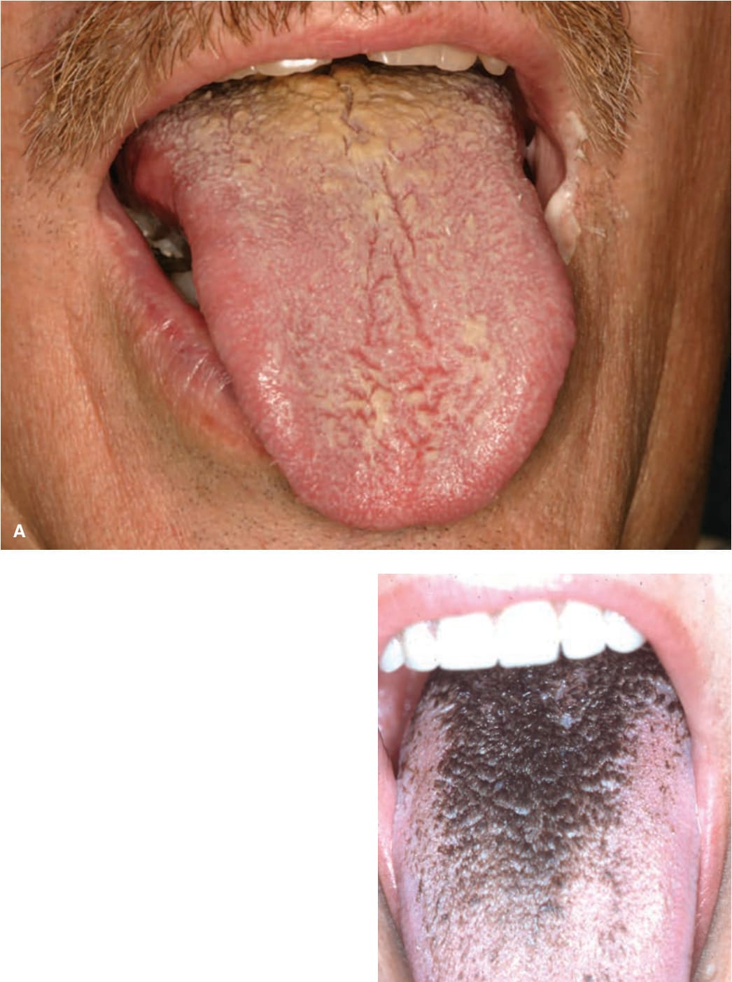
FIGURE 33-3 • (A) Hairy tongue Defective desquamation of filiform papilla noted in posterior aspect of tongue. Tongue has a white surface caused by retained keratin. (Used with permission from Dr. Nathaniel Treister.) (B) Black hairy tongue In this example, chromogenic bacteria have stained the tongue black.
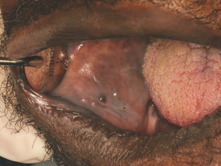
FIGURE 33-12 • Leukoedema In this variant of normal, there is bluish and whitish discoloration of mucosa that blanches when the cheek is stretched. (Used with permission from Dr. Nathaniel Treister.)
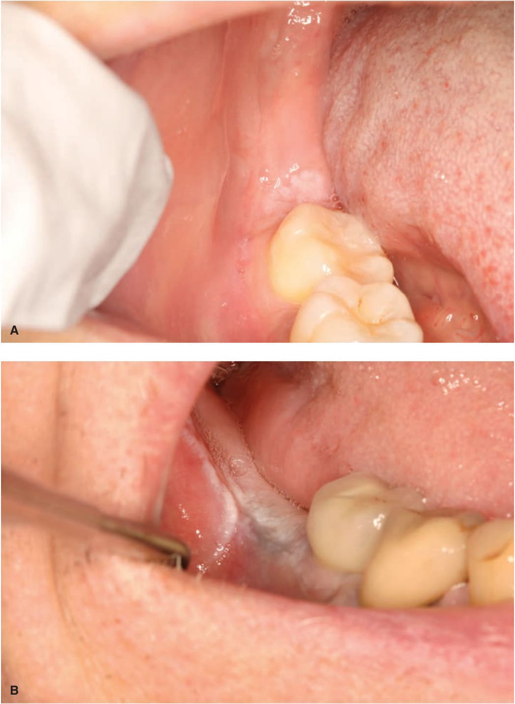
FIGURE 33-13 • (A, B) Lichen simplex chronicus Note the white plaque in the retromolar pad (after third molar extractions). These are often seen on edentulous ridge after extractions. (Used with permission from Dr. Sook-Bin Woo.)
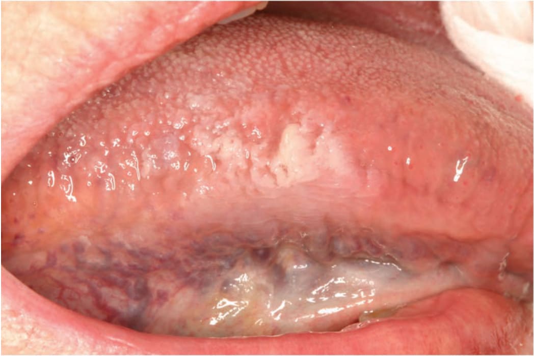
FIGURE 33-14 • Chronic chewing A wedge-shaped white papule is noted on the lateral surface of the tongue. (Used with permission from Dr. Sook-Bin Woo.)
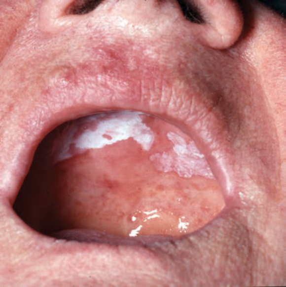
FIGURE 33-15 • Nicotine stomatitis Posterior palate shows erythematous pinpoint papules at sites of ducts and white patches where chemical irritation has caused chronic inflammation.
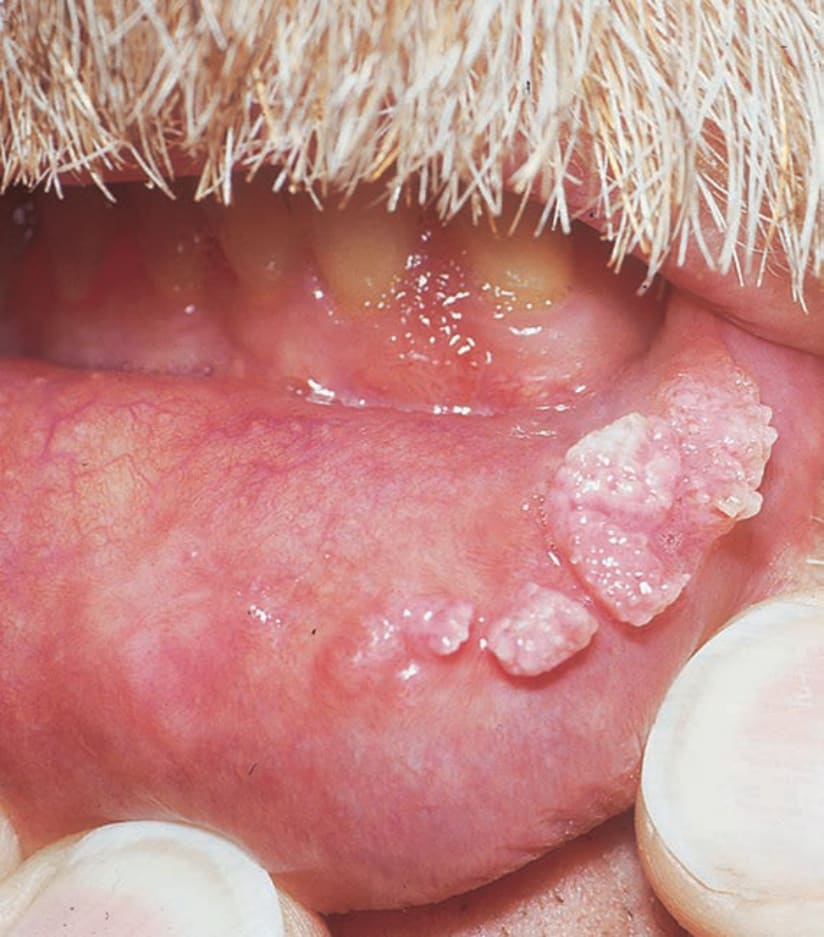
FIGURE 33-16 • Condyloma acuminatum: Mucosal lip Cluster of white cauliflower-floret-like lesions on the mucosa of the lower lip.
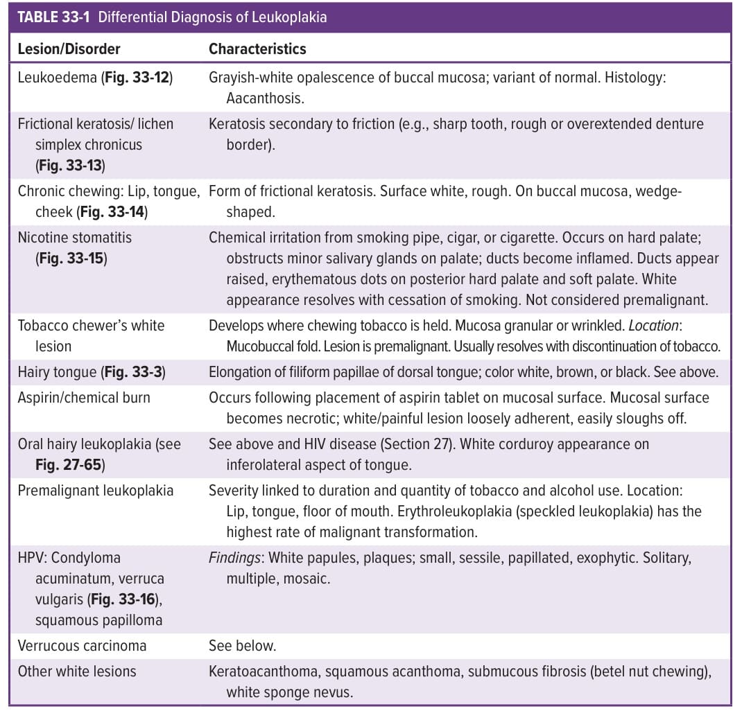
TABLE 33-1 Differential Diagnosis of Leukoplakia